| In EELS measurements, the zero-loss drift, spectrum drift and energy drift in EELS measurements originates from three main sources:
- High-Frequency Jitter:
- Origin: This type of energy shift is caused by electronic instabilities, such as noise or fluctuations in the power supply or other electronic components of the microscope (refer to page2585).
- Impact: High-frequency jitter can introduce small, rapid fluctuations in the energy scale of the EELS spectrum. However, these fluctuations are typically considered negligible, especially for core-loss EELS data, which is acquired at lower energy dispersion settings (meaning the energy resolution is higher, and the impact of jitter is less significant).
- The high-frequency jitter is apparent only in the low-loss data.
- Energy-Loss Deviation (Chromatic Correction):
- Origin: This deviation arises from systematic variations in the angle at which the electron beam enters the spectrometer as it scans across different positions on the sample. These variations can lead to changes in the energy loss spectrum depending on the probe position.
- Impact: Known as chromatic aberration or chromatic correction, this effect can cause the measured energy loss to vary slightly across the sample, leading to inaccuracies in the energy calibration of the EELS data. Correcting for this is important for obtaining precise and accurate measurements.
- This energy drift is normally the dominant contributor.
- Time-Dependent Energy Drift:
- Origin: This drift occurs over the duration of the experiment and is usually caused by slowly varying environmental factors such as temperature changes in the microscope or its surroundings.
- Impact: Thermal drift can induce energy shift. Thermal drift refers to the slow movement of the sample due to temperature changes during the acquisition process. As the sample drifts, the relative position of the electron beam and the sample can change, leading to variations in the recorded energy of the electrons. This effect can cause an apparent energy shift in the spectra, which needs to be corrected to ensure accurate data interpretation. Over time, this drift can cause a gradual shift in the energy scale of the EELS spectrum, leading to potential inaccuracies in the energy measurement if not corrected. This is a common issue in longer acquisitions, where temperature variations and other environmental factors can subtly alter the energy calibration.
These three sources of energy shift can affect the accuracy and precision of EELS measurements, and understanding and correcting for them is crucial in high-resolution EELS analysis. The energy-shift artifacts can normally be corrected to
within 1–2 eV.
It was proposed that the energy drift from low-frequency instabilities can be corrected by software techniques [1, 2], while the energy drift from high-frequency energy instabilities can be corrected by high speed acquisition technique [3].
A limitation of the magnetic prism-based EELS detection system is the spectrum drift in the energy dispersion direction. Various sources of instability in the microscope contribute to this effect: high voltage fluctuation, magnetic field creep etc. This places a fundamental limit on the useful exposure time. Furthermore, experimental efforts in electron spectroscopy consist largely in reducing unexpected electrical noise and specimen drift.
In order to improve the energy resolution of EEL spectrum, before spectrum deconvolution, scripts (e.g. applied in Gatan Digital Micrograph) can be used to automatically acquire and store each spectrum separately, and then to evaluate and correct the energy drift in each acquisition. [4, 5] After the drift correction, the EEL spectrum can be deconvoluted using software. For instance, some deconvolution techniques are Fourier ratio method, maximum-entropy (ME) [6,7] and Richardson–Lucy (RL) algorithms [8].
Examples in Figure 4891 shows the effects of energy resolution enhanced by energy-drift correction and deconvolutions in the EEL spectrum of h-BN. The EEL spectra are acquired with an exposure time of 80 ms, a probe current of 100 pA and a high energy-dispersion (0.021 eV ch–1). Figure 4891 (a) shows a blind-sum spectrum with a wide energy spread of 0.48 eV in FWHM (full width at half maximum) due to the energy drift during data acquisition. Figures 4891 (b) shows the improvement by the energy-drift correction, reflecting the inherent high energy-resolution of a cold field emission electron gun (CFEEG). Figures 4891 (c) and (d) show the boron K-edge spectra before and after drift correction, respectively, with π* peak reduced from 1 to 0.52 eV. Figures 4891 (e) and (f) shows further improvement by RL (Richardson-Lucy) and ME (maximum-entropy) deconvolution, respectively.
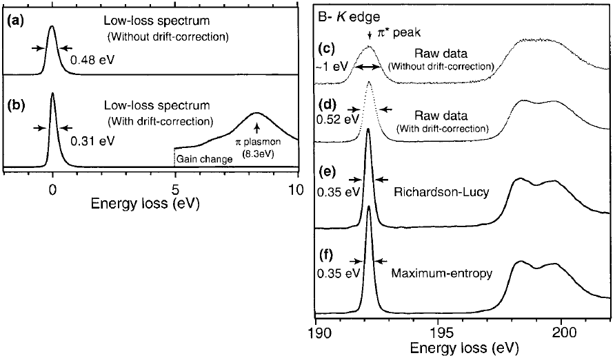
Figure 4891. Low-loss and core-loss spectra of h-BN. (a) and (c) raw spectrum, (b) and (d) spectra after drift correction, and (e) and (f) deconvoluted B K-edge ELNES using RL algorithm and ME algorithm, respectively.
Adapted from [5]
[1] Kimoto, K., Matsui, Y., 2002. Software techniques for EELS to realize
about 0.3 eV energy resolution using 300 kV FEG-TEM. J. Microsc.
208, 224–228.
[2] Kimoto, K., Ishizuka, K., Mizoguchi, T., Tanaka, I., Matsui, Y., 2003. The
study of Al-L23 ELNES with resolution-enhancement software and firstprinciples
calculation. J. Electron Microsc. 52, 299–303.
[3] Koji Kimoto, Kazuo Ishizuka, Toru Asaka, Takuro Nagai, Yoshio Matsui, 0.23 eV energy resolution obtained using a cold field-emission gun and a streak imaging technique, Micron 36 (2005) 465–469.
[4] Kimoto K and Matsui Y (2002) Software techniques for EELS to realize
about 0.3 eV energy resolution using 300 kV FEG-TEM. J. Microsc.
208: 224–228.
[5] Koji Kimoto, Kazuo Ishizuka, Teruyasu Mizoguchi, Isao Tanaka and Yoshio Matsui, The study of Al-L23 ELNES with resolution-enhancement software and first-principles calculation, Journal of Electron Microscopy 52(3): 299–303 (2003).
[6] Kuzuo R and Tanaka M (1993) Resolution enhancement of electron
energy-loss spectra using the maximum entropy method. J. Electron
Microsc. 42: 240–243.
[7] Overwijk M H F and Reefman D (2000) Maximum-entropy deconvolution
applied to electron energy-loss spectroscopy. Micron 31: 325–331.
[8] Gloter A, Douiri A, Tencé M, Imhoff D, and Colliex C (2002) Improving
energy resolution of EELS spectra: an alternative to the monochromator
solution. In: Proc. of 15th ICEM, Durban, South Africa, pp. 141–142.
|
