| |
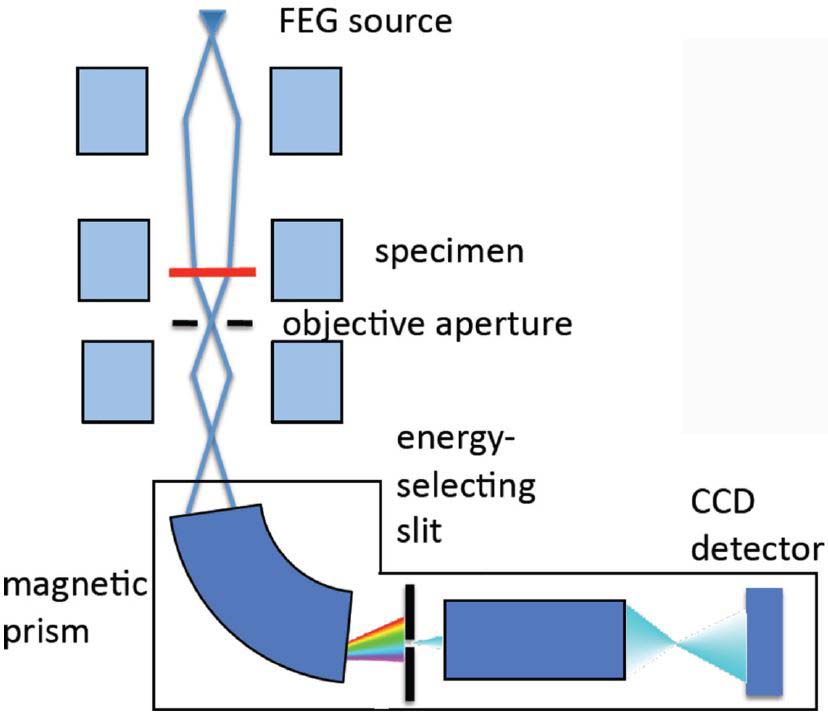
EFTEM |
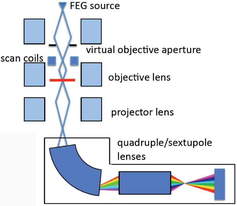
EELS Mapping in STEM Mode |
| |
=============================== |
=============================== |
| |
|
|
Electron beam |
Large, parallel, and fixed-beam illumination in TEM mode |
A focused beam is scanned across the area of interest of the specimen in STEM mode |
Acquired data |
The core-loss processes probed by EELS and
EFTEM are identical |
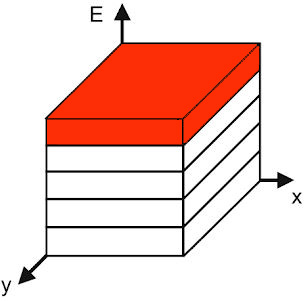
Acquires energy-selected images with spectral information integrated over a specific energy-loss range as defined by an energy-selecting slit |
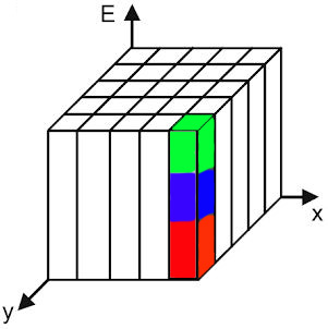
Acquires spectral data at each pixel in a serial manner |
| Image is recorded in parallel so that an energy-filtered image or an elemental map can be obtained in several seconds or a few minutes |
The data are acquired one pixel at a time in a serial mode |
|
|
|
|
Spatial information |
Allows spatial information to be acquired in parallel, with spectral information captured
serially |
Spectral information is
acquired in a spatially serial manner, requiring multiple acquisitions to build up a spatial picture |
Illumination angle |
Small: similar to CTEM |
Large in order to optimize the probe size and current |
Collection angle |
Small: similar to CTEM |
Large in order to optimize the collection
efficiency |
Spatial resolution |
With delocalization effects providing the fundamental limiting factor |
| Lower because
both the illumination and EELS collection angles are small |
Considerably higher because
both the illumination and EELS collection angles are large |
Spectral resolution |
Poorer |
Better |
Aberration effect |
More aberration effects |
In the STEM, the
EELS collection optics does not focus the electrons into an image and
thus is much less affected by aberrations [1] |
Spectral information |
Is probed serially by acquiring a number of images separated in energy loss |
Acquires spectral data at each pixel
in a serial manner |
Acquisition efficiency |
Less efficient since only the energy region defined by the energy-selecting slit is recorded |
More efficient since the information at each pixel is recorded |
Specimen damage |
More radiation damage since the total electron dose is more |
Less radiation damage since the total electron dose is less |
Data acquisition time |
Short since the recording time is almost independent of the number of pixels due to spatially parallel nature and larger total electron dose
|
Long due to non-negligible readout time per spectrum |
Dose-rate-dependent specimen damage |
Minimized |
There are such damages, e.g. hole-drilling and movement of segregants |
[1] Stephen J. Pennycook, Peter D. Nellist, Scanning Transmission Electron Microscopy Imaging and Analysis, 2011.
[2] M.A. Aronova and R.D. Leapman, Development of Electron Energy Loss Spectroscopy in the Biological Sciences, MRS Bull. 2012, 37(1): 53–62. doi:10.1557/mrs.2011.329.