=================================================================================
For EELS analysis, the final energy resolution of the system is limited by the energy spreads of the beam at the electron source and due to the spectrometer optics. The resulting “degraded” spectrum is given by,
 ------------------------------- [2637] ------------------------------- [2637]
where,
H -- The acquired spectrum,
W -- The true (non-degraded) spectrum,
p -- The Point Spread Function (PSF) of the system.
Note that a monochromator for the electron source or data deconvolution is necessary in the frontiers of TEM-EELS if the energy spread of the available electron source in TEM is larger than the intrinsic fine structures of spectra.
Comparing to using monochromation system, a less costly method to improve energy resolution is mathematical deconvolution. This process utilizes an experimentally determined function which represents the inherent energy spread of the electron optics system in the EM.
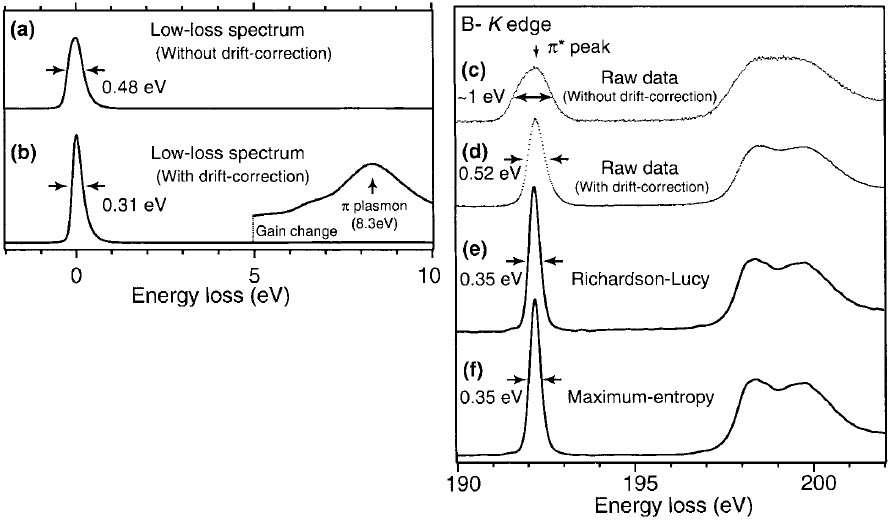
For instance, Nelayah et al [1] have demonstrated the successful application of the Richardson-Lucy deconvolution algorithm to improve the energy solution acquired from silver nanoprisms. More examples in Figure 2637 shows the effects of energy resolution enhanced by energy-drift correction and deconvolutions in the EEL spectrum of h-BN. The EEL spectra are acquired with an exposure time of 80 ms, a probe current of 100 pA and a high energy-dispersion (0.021 eV ch–1). Figure 2637 (a) shows a blind-sum spectrum with a wide energy spread of 0.48 eV in FWHM (full width at half maximum) due to the energy drift during data acquisition. Figures 2637 (b) shows the improvement by the energy-drift correction, reflecting the inherent high energy-resolution of a cold field emission electron gun (CFEEG). Figures 2637 (c) and (d) show the boron K-edge spectra before and after drift correction, respectively, with π* peak reduced from 1 to 0.52 eV. Figures 2637 (e) and (f) shows further improvement by RL (Richardson-Lucy) and ME (maximum-entropy) deconvolution, respectively.

Figure 2637. Low-loss and core-loss spectra of h-BN. (a) and (c) raw spectrum, (b) and (d) spectra after drift correction, and (e) and (f) deconvoluted B K-edge ELNES using RL algorithm and ME algorithm, respectively.
Adapted from [2]
[1] Nelayah, J. et al. Mapping surface plasmons on a single metallic nanoparticle. Nature Phys. 3, 348-353 (2007).
[2] Koji Kimoto, Kazuo Ishizuka, Teruyasu Mizoguchi, Isao Tanaka and Yoshio Matsui, The study of Al-L23 ELNES with resolution-enhancement software and first-principles calculation, Journal of Electron Microscopy 52(3): 299–303 (2003).
|
