=================================================================================
Examples of pole piece gaps in the objective lenses in EMs are HD 2700C SEM/STEM has a 3.8 mm gap and HD 2300A SEM/STEM has 5 mm gap.
Figure 2943 shows the schematic illustration of top-entry specimen stage together with the top-entry lens in TEMs. The magnetic gap (pole piece gap) is also indicated.
Figure 2943. Schematic illustration of top-entry specimen stage together with the top-entry lens.
TEMs with side-entry specimen stages usually requires a wider objective lens gap which degrades the instrument's analytical properties.
Comparing with X-ray crystallography, the major difficulties in electron diffraction technique in TEM are smaller tilt range with goniometer stages and smaller physical dimension of specimens placed in the objective lens polepiece gap.
The focusing properties of a magnetic lens may be given in terms of a parameter k2,
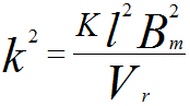 ---------------------------------------- [2943] ---------------------------------------- [2943]
where,
Bm -- The maximum value of the magnetic induction between the polepieces.
l -- A length characteristic of the lens, which can be the half-width of the axial magnetic field distribution, or the polepiece gap S or the radius R of the bore through polepieces.
K -- A numerical factor incorporating the charge and mass of the electron.
Vr -- The relativistically corrected value of the electron beam voltage.
To minimize the aberrations, the objective lens has to be strong. Therefore, Bm must be kept as high as possible but without saturating the soft iron polepieces, while l should be kept small in order to keep the lens compact and the whole microscope column reasonably short. Assuming k2 maintains constant as the accelerating voltage is raised, with Bm fixed, l would have to be increased in proportion to (Vr)1/2.
|