=================================================================================
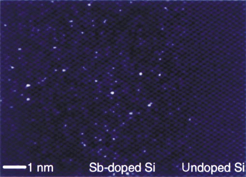
Voyles et al [1] analyzed Sb (antimony) doped Si (silicon) in the <110> zone-axis orientation using ADF-STEM (annular dark-field scanning transmission electron microscopy) operated at 200 kV, directly observed individual Sb atoms at atomic-resolution, and identified the Sb clusters in Si responsible for the saturation of charge carriers. The size, structure, and distribution of these clusters were determined with a Sb-atom detection efficiency of almost 100%. The brightest dots on the left in the ADF-STEM image in Figure 3511a are atomic columns containing at least one Sb atom and the undoped region on the right shows no such very bright dots. Regarding the quality of the TEM sample, their simulations showed that for a Si column containing a single Sb atom to be more than 25% brighter than a pure Si column, the sample must be less than 50 Å thick. Random variations in thickness must be less than the contrast of one Sb atom, and the surfaces must be free from native amorphous SiO2 layers and amorphized bulk Si layers.

Figure 3511a. ADF-STEM image of a cross section of highly Sb-doped Si.
Figure 3511b shows high-resolution HAADF Z-contrast STEM images presenting the interface between Si3N4 matrix (on the top of each image) and the mixtures (at the bottom of each image) of Yb2O3 and Al2O3 (a) and Yb2O3 and SiO2 (b). The Yb atoms, visible here as bright spots due to enhanced electron scattering (giving higher Z-contrast), are located periodically along the interface. The images are oriented along the low-index zone axis [0001] of Si3N4. Note the hexagonal ring structures represent the typical Si3N4 atomic structure [3] based on phase reconstruction microscopy techniques, even though the individual close atomic positions of Si–N cannot be fully resolved here because the limit of the spatial resolution of the STEM system used.
![High-resolution STEM images showing the atomic interface between a Si3N4 matrix grain, oriented along the low-index zone axis [0001], and two different mixtures](image1/3445.jpg)
Figure 3511b. High-resolution STEM images showing the atomic interface between a Si3N4 matrix
grain and two different mixtures. Adapted from [2]
[1] P. M. Voyles, D. A. Muller, J. L. Grazul, P. H. Citrin, and H.-J. L. Gossmann, Atomic-scale imaging of individual dopant atoms and clusters in highly n-type bulk Si, Nature, 416 (2002) 826.
[2] A. Ziegler, M. K. Cinibulk, C. Kisielowski, and R. O. Ritchie, Atomic-scale observation of the grain-boundary structure of Yb-doped and heat-treated silicon nitride ceramics, Appl. Phys. Lett. 91, 141906 (2007).
[3] A. Ziegler, J. C. Idrobo, M. K. Cinibulk, C. Kisielowski, N. D. Browning, and R. O. Ritchie, Appl. Phys. Lett. 88, 041919 (2006).
|