=================================================================================
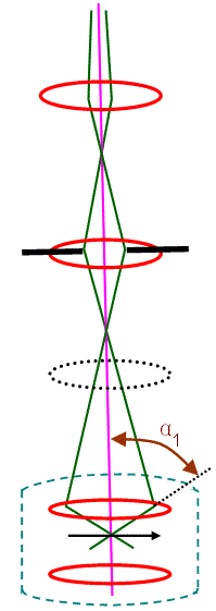
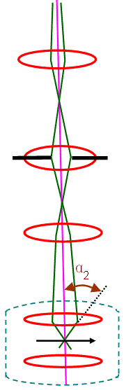
The schematic illustrations in Figure 3705a show the convergent illumination configurations of various modes in TEMs. In the CTEM condition in Figure 3705a (a), the condenser mini-lens (CM lens) is strongly excited, and incident electrons are focused on the pre-focal point of the objective pre-field, resulting in a parallel illumination on a wide area on the specimen and providing highly coherent electron illumination. In the EDS condition in Figure 3705a (b), the CM lens is turned off and the incident electrons are focused on the specimen by the objective pre-field, resulting in a small-probe illumination. In this case, the illumination angle (α1) is large so that high beam intensity is obtained for a small area in the analytical EDS method. In the NBD mode in Figure 3705a (c), a smaller condenser aperture is used to form a smaller illumination angle (α2). Therefore, a small-diameter probe with relatively high coherence in the illumination is achieved. In the illumination condition in Figure 3705a (c), the illumination angle (α) with a constant probe size can be changed by changing the excitations of the condenser lenses and the CM lens to obtain the incident illumination to form ideal convergent beam electron diffraction (CBED) patterns.
Figure 3705a. Convergent illumination configurations: (a) CTEM mode, (b) EDS mode and (c) NBD mode.
Figure 3705b gives a very simplified illustration of how the incident electrons travel through the TEM column. r is the real space coordinates in the sample or image, u is the reciprocal space coordinates and θ is the scattering angle.
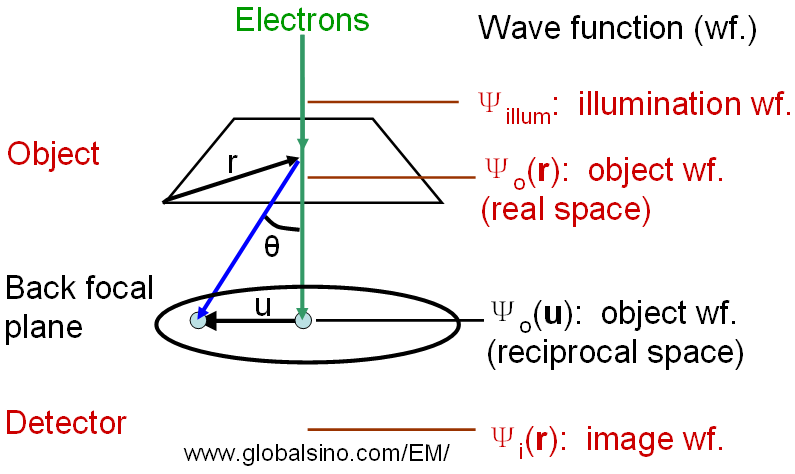
Figure 3705b. Simplified illustration of how an image is formed in a TEM column.
|