Image Sensors/Electronic Optical Imaging Sensors - Practical Electron Microscopy and Database - - An Online Book - |
|||||
| Microanalysis | EM Book https://www.globalsino.com/EM/ | |||||
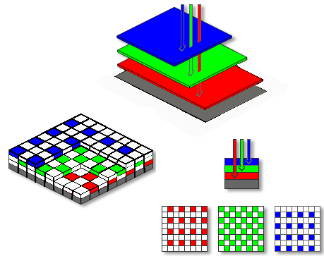
An electronic optical imaging sensor consists of photonsensitive regions which are called pixels. Each pixel converts photons into electrons according to its incident light intensity, accumulates the generated electrons (charge collection) according to its well capacity during the exposure time, and transfers the collected electrons via a charge-to-voltage converter according to its bit depth and readout mechanism. Each of these processes, charge generation, collection, and transfer is also accompanied with various types of noise, which affects the quality of formed image. For instance, a dark current noise is caused by thermal effects during the exposure time and generates nonphotonic charges in the wells. The readout noise is added to the readout signal simultaneously while the collected charge is transferred from pixel to the on-chip amplifier and then, is converted into an analog voltage. The cone-shaped cells inside human eyes are sensitive to red (R), green (G), and blue (B) which are the "primary colors". All other colors are combinations of these primary colors. In the conventional photography, the red, green, and blue components of light expose the corresponding chemical layers of color films. The new Foveon sensors are based on the same principle, and have three sensor layers that detect the primary colors as shown in Figure 3975. In the color digital imaging, a mosaic of square tiles or "pixels" of uniform color basically are so tiny that it appears uniform and smooth to our eyes.
Figure 3975. Principle of Foveon sensor.
|
|
||||