=================================================================================
Much of the spectral information obtainable from EELS is similar to that given by synchrotron-XAS (x-ray absorption spectroscopy), so that EELS in TEM has been referred to as a synchrotron in electron microscopes [1]. The electric field of an incident high energy x-ray can eject an electron of condensed matter. Page4777 lists the comparison between XAS and EELS. One important difference from EELS is that x-rays transfer all their energy by ejecting electrons from their initial state in the material.
Irradiating by the x-ray, the electrons in the sample feel a force parallel to the polarization, resulting in a change of momentum. This force is due to a coupling between the electron and the x-ray (of angular frequency ω), given by,
 ----------------------------- [4778a] ----------------------------- [4778a]
where,
ε -- Unit vector specifying the polarization of the x-ray;
d -- Dipole operator, given by;
E0 -- Magnitude of the x-ray electric field.
 ----------------------------- [4778b] ----------------------------- [4778b]
where,
-e -- Charge of the electron.
The cross-section for x-ray absorption from the ground state |ψ0> of the Hamiltonian H0 by applying Fermi's golden rule can be given by, [2]
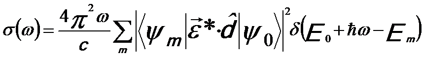 ----------------------- [4778c] ----------------------- [4778c]
or,
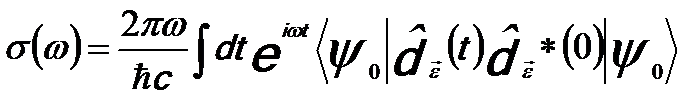 ----------------------- [4778d] ----------------------- [4778d]
 ----------------------- [4778e] ----------------------- [4778e]
where,
ħ -- Reduced Planck constant;
c -- Speed of light.
The most common way of performing XAS is to let x-rays penetrate a thin foil with typical thickness of 10 - 100 µm and then measure the fraction of the incident beam intensity penetrating the foil as a function of the energy of the incident x-rays. Therefore, an x-ray source, such as synchrotrons, that is intense over a large energy range is needed. For energies below an absorption edge, the x-rays penetrate rather easily without absorption because the Pauli exclusion principle prevents excitation, while above the absorption edge threshold the x-rays have sufficient energy to eject core electrons to empty states above the Fermi level.
X-ray absorption extended fine structure (EXAFS) analysis generally uses inner-shell edges with binding energies of order 10 kV that provide a range of 1000 - 2000 eV of fine structure information. EXAFS is particularly useful for amorphous and highly disordered materials.
The first use of density functional theory (DFT) for the calculation of X-ray absorption spectra was done by Müller et al. using a linearized augmented plane waves method in the late 70s [3].
[1] Brown L M 1997 A synchrotron in a microscope Proc. EMAG97
(Cambridge) (Inst. Phys. Conf. Ser. 153) pp 17–21
[2] Fermi, E., Nuclear Physics. University of Chicago Press, 1950.
[3] Müller, J.E., Jepsen, O., 1978. Systematic structure in the K-edge photoabsorption
spectra of the 4d transition metals: theory. Phys. Rev. Lett. 40 (11), 720 -
722.
|