=================================================================================
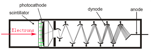
In electron microscopes, combination of YAG scintillator and photomultiplier (PM) is used in some electron detectors as shown in Figure 4865a. The
generated visible light in scintillator travels via fiber optics to a photocathode. The light is re-converted to electrons at the photocathode. The electron signal is
multiplied by several dynodes (electrodes) in the PM vacuum tube before being
used to modulate the display screen or electron detectors.

Figure 4865a. Schematic of scintillator-photomultiplier system.
We can have many benefits from the combination of scintillator-PM systems. First, a plastic scintillator allows pulse counting up to several MHz. Second, due to the high detective quantum efficiency (DQE), the gain of the scintillator-PM system is very high and is in the order of 10n. Here, n is the number of dynodes (normally n ≥ 8). But the gain of semiconductor detector is about 104. Third, we can benefit from the high signal-to-noise ratio and wide bandwidth in a scintillator. The signal-to-noise ratio in the scintillator is very high and is better than that of
semiconductor detectors. The bandwidth of the
scintillator is in the MHz range. Therefore, it is easy to obtain
low-intensity images as well as TV-rate images, and thus, we even though can capture data at room illumination instead in the dark room and record dynamic in-situ events.
Comparing with semiconductor detectors, the weakness of the scintillator-PM system are mainly 1) Scintillators are more susceptible to radiation damage and must be changed or mechanically shifted from time to time; 2) Scintillator-PM systems are more expensive and bigger; 3) Scintillators hae rather low electron-to-counts conversion efficiency and low energy-conversion efficiency ( between ~ 2% and ~20%); 4) It may be necessary to correct for response nonlinearity for count rates > 1 MHz [1].
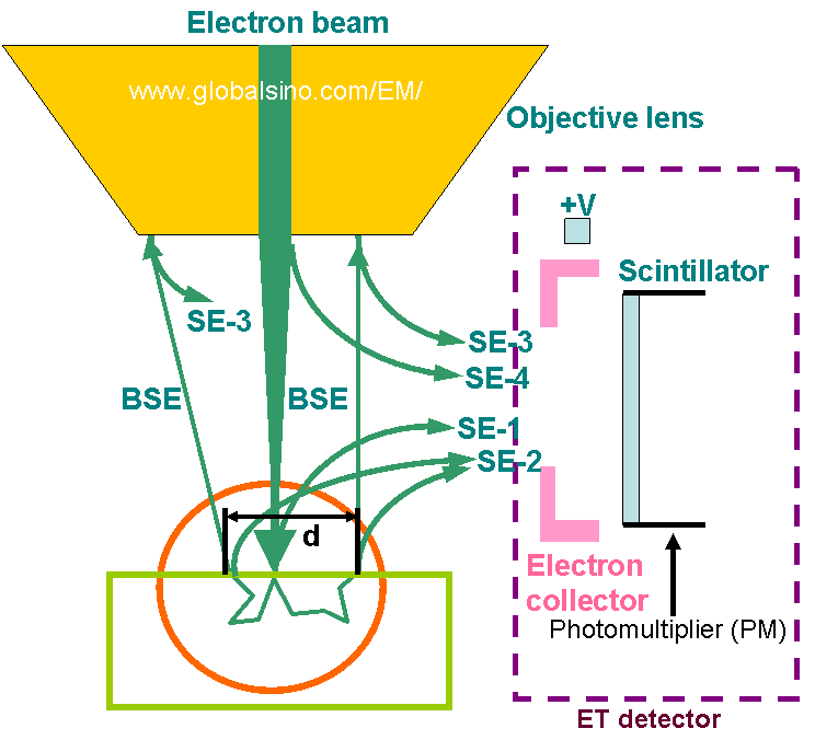
Figure 4865b shows an example of scintillator-photomultiplier systems used in Everhart-Thornley (ET or E-T) detectors installed in most scanning electron microscopes (SEM). This type of systems is mainly used for detection of secondary electrons (SEs).

Figure 4865b. Three types of secondary electrons collected by the Everhart-Thornley (ET) detector formed
by scintillator-photomultiplier systems. Adapted from Reimer [2].
[1] R. F. Egerton, Y.-Y. Yang, and S. C. Cheng, “Characterization and use of
the Gatan 666 parallel-recording electron energy loss spectrometer”. Ultramicroscopy
48, 239 (1993).
[2] Reimer, L., Scanning Electron Microscopy, Springer Verlag, New York, 1985.
|

