Techniques used to Detect/Measure Charges in EM Samples - Practical Electron Microscopy and Database - - An Online Book - |
||||||||
| Microanalysis | EM Book http://www.globalsino.com/EM/ | ||||||||
| ================================================================================= | ||||||||
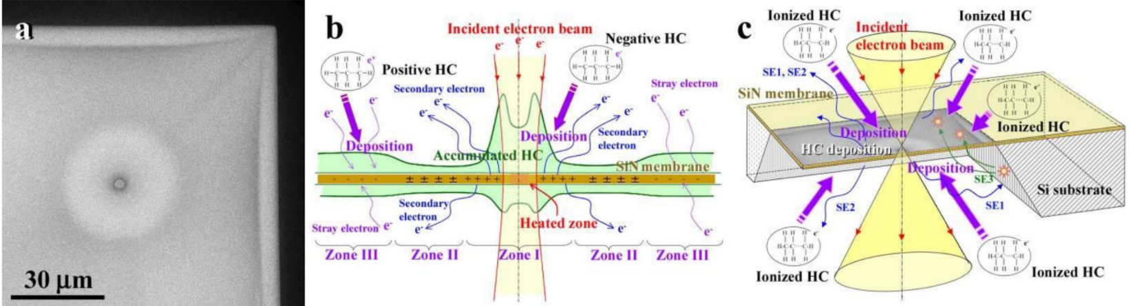
To detect charge distributions on insulating films, a hydrocarbon (HC) replica method had been applied [1] and indicated the distribution of absorption and accumulation of positively or negatively ionized hydrocarbon molecules in residual gases in a TEM column [2, 3]. Figure 999 shows a hydro-carbon contamination layer formed on a Si3N4 membrane in thickness of 30 nm. The accelerating voltage of the electron beam used to irradiate on the Si3N4 membrane was 75 kV. The charge distributions consist of three zones around the beam irradiation point at the center:
[1] Ken Harada, Keiko Shimada, Kodai Niitsu, Teiji Katsuta, Teruaki Ohno, and Daisuke Shindo1, Transmission Electron Microscope Observation of Charge Distribution on Insulating Thin Films by Hydro-carbon Deposition, Microsc. Microanal. 23 (Suppl 1), 2017.
|
||||||||
| ================================================================================= | ||||||||
|
|
||||||||