=================================================================================
Figure 1945a shows the TEM images of 1-nm He (helium) bubbles in palladium tritides when the specimen is oriented far from any Bragg conditions of the matrix of the palladium tritides [1]. For negative defocus (underfocus), the bubbles appear as white dots surrounded by a dark fringe, while for positive defocus (overfocus), the dots are black with white fringe.
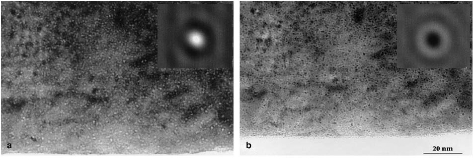
Figure 1945a. The TEM images of 1-nm He (helium) bubbles in palladium tritides: (a) Underfocus and (b) Overfocus. The insets present theoretically simulated TEM contrasts of centered He bubbles in 6.5 nm thick TEM specimen. Adapted from [1]
Figure 1945b shows a HRTEM image of Bi12.8Co0.2Mo5O34±δ crystal along [010] zone axis. The intense white reflections correspond to voids in the crystal structure which was confirmed by simulation.
![HRTEM image of Bi12.8Co0.2Mo5O34±δ along [010] zone axis](image2/1945b.GIF)
Figure 1945b. HRTEM image of Bi12.8Co0.2Mo5O34±δ along [010] zone axis. In the simulated inset, the spheres denote Bi and O atoms, tetrahedra denote MoO4 units (simulation condition: defocus = −17 nm and thickness = 4.7 nm). [2]
[1] S. Thiébaut, B. Décamps, J. M. Pénisson, B. Limacher, A. Percheron Guégan, TEM study of the aging of palladium-based alloys during tritium storage, Journal of Nuclear Materials 277 (2000) 217-225.
[2] Z. A. Mikhailovskaya, E. S. Buyanova, S. A. Petrova, M. V. Morozova, V. M. Zhukovskiy, R.G. Zakharov, N. V. Tarakina, I. F. Berger, Cobalt-doped Bi26Mo10O69: Crystal structure and conductivity, Journal of Solid State Chemistry 204 (2013) 9–15.
|