=================================================================================
Figure 1950 shows a schematic of a focused, conical electron beam as it strikes the surface of the specimen, showing over-focus, in-focus and under-focus. For the over-focus case, the magnetic field is too strong, and the beam converges to a crossover above the surface of the specimen; however, for the under-focus case, the magnetic field is too weak so that the beam is not fully brought to crossover before it strikes the surface of the specimen. For both over-focus and under-focus cases, the beam diameter is broader than optimal, resulting in an out-of-focus image.
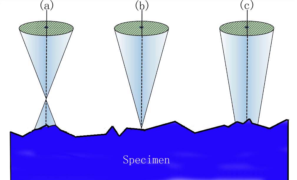
| Figure 1950. Schematic of a focused, conical electron beam as it strikes the surface of the specimen, showing over-focus (a), in-focus (b), and under-focus (c). |
Note that unlike the experience with light microscopy, underfocus TEM images often appear somewhat sharper than in-focus electron images.
|