|
This book (Practical Electron Microscopy and Database) is a reference for TEM and SEM students, operators, engineers, technicians, managers, and researchers.
|
=================================================================================
Figure 2933 shows seven (100) SrTiO3 high-resolution ADF STEM images. These images were obtained at a convergence angle of 28 mrad in the defocus range between -36 nm and +36 nm. It can be seen that the contrast reduces as a function of defocus.
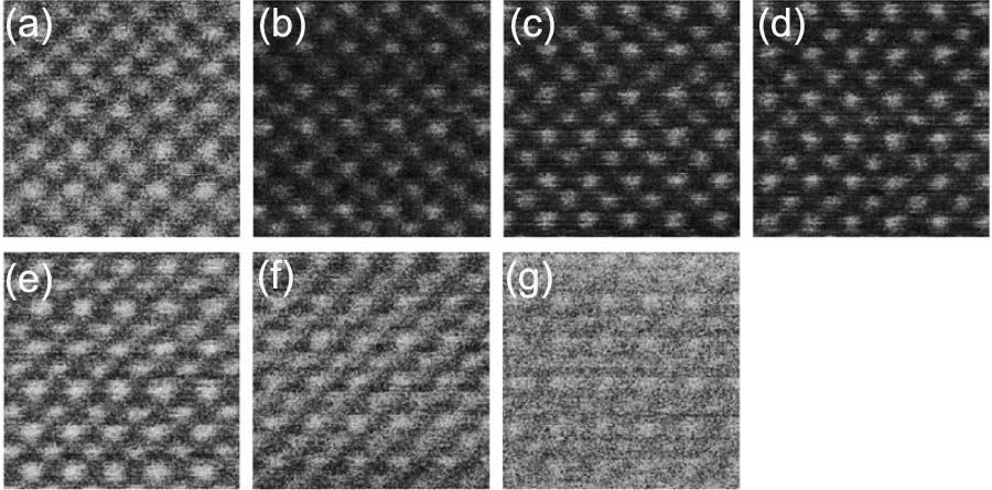
Figure 2933. ADF STEM images taken at different defoci of (a) -36, (b) -24, (c) -12, (d) 0, (e) +12, (f) +24, and (g) +36 nm. "-" represents under focus, while "+" represents over focus. Adapted from [1]
Based on the optical reciprocity theorem [2], BF-STEM images can be related to the HRTEM images of an atom as a phase object. Like HRTEM images, BF-STEM images are very sensitive to the focal conditions, and contrast reversal is often observed because of multiple scattering and phase shift due to defocus condition of the lens. A full interpretation of BF-STEM images can be done with phase contrast simulation in the same way as the HRTEM images. [3]
[1] H. Inada, D.Su, R. F. Egerton, M.Konno, L.Wu, J.Ciston, J.Wall, Y.Zhu, Atomic imaging using secondary electrons in a scanning transmission electron microscope: Experimental observations and possible mechanisms, Ultramicroscopy 111(2011)865–876.
[2] Born M, Wolf E. Principles of optics. Cambridge, UK: Cambridge University Press; 1997.
[3] Spence JCH. High resolution electron microscopy. 3rd ed. Oxford: Clarendon Press; 2003.
|