=================================================================================
Figure 3832a shows the generation of secondary electrons (SEs) and kinetic energies of the emitted SEs. EKE1 and EKE2 represent the kinetic energies of the two generated SEs. The kinetic energy of the generated SEs is normally in the range of 0 to 50 eV. ΔE1 and ΔE2 represent the energy losses of the incident electrons after the incident electrons interact with the electrons in the K and L3 subshells, respectively. E1 and E2 are the binding energies of the two electrons. E0 is the energy of the incident electrons in the EMs.
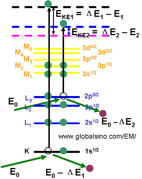
Figure 3832a. The generation of secondary electrons (SEs) and kinetic energies of the emitted SEs.
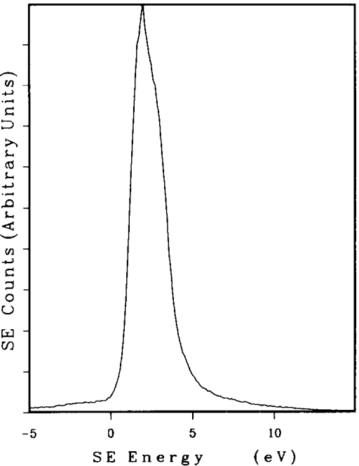
As shown in Figure 3832b, the SE (secondary electron) energy spectroscopy, which is the relative SE number as a function of the SE energy (N(E)), is characterized by the most probable energy (eVmp) and the full width at half maximum (FWHM). The spectroscopy generally varies in a range with the composition of the materials and surface condition.
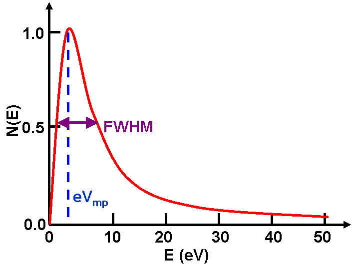
Figure 3832b. Schematic illustration of SE (secondary electron) energy spectroscopy.
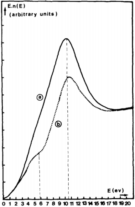
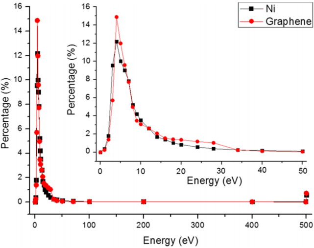
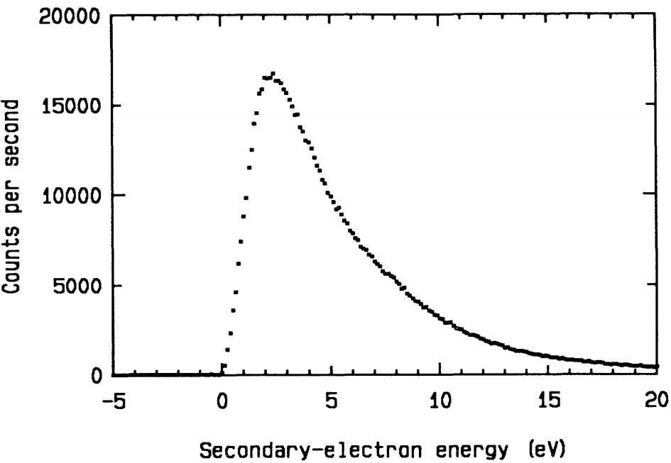
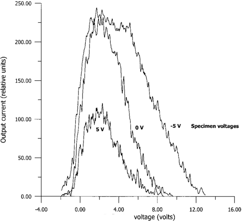
Figure 3832c shows some examples of secondary electron spectra obtained from different materials.
|
 |
(a) |
(b) |
 |
|
(c) |
(d) |
|
|
(e) |
|
Figure 3832c. Secondary electron spectra obtained from different materials: (a) MgO smoke crystal [1], (b) thin amorphous carbon film [2], (c)
metal coat biased at voltages of -5, 0, and +5 V [3], (d)  sputtered clean Al and sputtered clean Al and  oxidized Al [4], and (e) Ni and graphene [5]. oxidized Al [4], and (e) Ni and graphene [5]. |
Note that secondary electrons may lose some of their energy by the bremsstrahlung process, in which the bremsstrahlung x rays carry energy away.
[1] J. Liu and J.M. Cowley, Imaging with high-angle scattered electrons and secondary electrons in the STEM, Ultramicroscopy, 37, 50 (1991).
[2] Folbert J. Pijper and Pieter Kruit, Detection of energy-selected secondary electrons in coincidence with energy-lass events in thin carbon foils, Physical Review B, 44, 17, 9192 (1991).
[3] A. Khursheed and N. Karuppiah, An add-on secondary electron energy spectrometer for scanning electron microscopes, Review of Scientific Instruments, 72, 3, 1708 (2001).
[4] D. Massignon, F. Pellerin, J. M. Fontaine, C. Le Gressus, and T. Ichinokawa, Comparison of the secondaryelectron spectrum with the electronloss spectrum on pure Al by lowenergy electronreflection spectroscopy, Journal of Applied Physics, 51, 808 (1980).
[5] Jialong He, Jie Yang, Yufei Peng, Jidong Long, Zhen Yang, Tao Wang, Ping Liu, Jie Li, Le Zheng, Pan Dong, Xi Li, Chaohui Lan, Wei Zhao, Erxiang Liu, and Jinshui Shi, Measurement of yield and spectrum of secondary electron emission and their characteristics under modification of conductive materials, Rev. Sci. Instrum. 90, 063304 (2019).
|