|
This book (Practical Electron Microscopy and Database) is a reference for TEM and SEM students, operators, engineers, technicians, managers, and researchers.
|
=================================================================================
In the arrangement of the basic imaging systems of conventional transmission electron microscopy (CTEM), an electron beam emitted by a source radiates the TEM specimen, usually through an illuminating system of lenses as shown in blue in Figure 4138a. The incident electrons interact with the specimen and thus are scattered. The electron beam after interaction is then focused by the objective lens, forming an image. The image is further magnified by projector lenses to produce an image at proper magnifications. Note that in modern TEM systems additional lenses, intermediate lenses are also placed between objective lens and projector lenses.
For scanning transmission electron microscopy (STEM), the same diagram can be used with a reversed direction of the electron beam also shown in Figure 4138a. The electron source for STEM replaces the TEM detection system (image plane). In modern STEM systems, a field-emission gun can be used to produce an electron source as small as 10 nm in diameter. One or more projector lenses in TEM (acting as condenser lenses in STEM) with a short focal length demagnify the source to provide the flexibility in the beam parameter. The beam is then demagnified further by the main lens in the systems, which is objective lens to form a small probe of 0.2 – 1 nm on the specimen. Therefore, a STEM probe can be considered as a demagnified image of the source. Note that the focal length of the objective lens can be as small as 1 mm. The scanning coils are deflector coils, built into the bore of the objective lens, and serve to scan the electron probe over the specimen. The detector on the STEM image plane collects the electrons transmitted, or scattered, by the specimen.
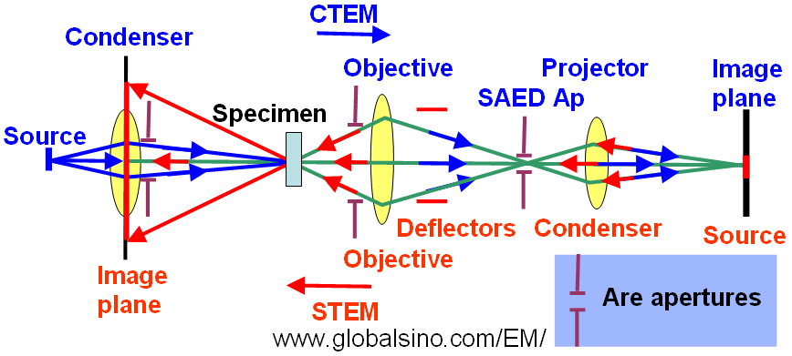
Figure 4138a. Schematic illustration of imaging geometries of TEM and STEM systems. The green lines show the common paths of the rays of CTEM and STEM illuminations. The blue arrows indicate the rays for the TEM illumination, while the red arrows indicate the directions of STEM rays. The STEM image is formed on the left-hand side, while the CTEM image is on the right-hand side. The yellow lenses are the common lenses for both STEM and CTEM systems.
Moreover, CBED (convergent-beam electron diffraction) patterns are obtained by producing an electron
probe of 5–20 nm in diameter as a demagnified image of the electron source (~10 nm - 40 µm).
Figure 4138b shows the structure of the electron probe-forming system in STEM mode in JEOL JEM-2010F TEMs. A setup, consisting of electrostatic gun lens and twin condenser lens system, controls the de-magnification of the Schottky field emission source. Compared with the cold field emitter, the Schottky electron source has a much larger emission area. A large de-magnification factor between the source and probe reduces this probe-broadening effect. In addition, the beam current decreases due to the probe-forming aperture. A cross-over is formed between the two condenser lenses (C1 and C2) by employing near-maximum excitation in the C1 lens, resulting in a large source de-magnification. The C2 lens and the gun lens are used to tune the probe coherence further by setting the probe size. In the STEM mode, both condenser and objective mini-lenses are tuned off.
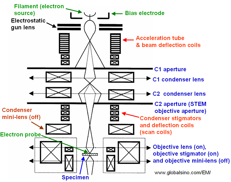
Figure 4138b. Schematic illustration of the probe-forming electron optics in STEM mode in JEOL JEM-2010F TEMs.
|