Table 1197a. Dependence of contrast in annular dark-field (ADF) images.
Factors |
| Specimen thickness [1,2] |
| Crystalline orientation [3] |
| Chemical composition [4] |
| Surface oxidation [5] |
| Inner detection angles [1,2] |
The ADF signal can be obtained by integrating the CBED intensity or other scattered intensity over the proper detector dimensions. The ADF intensity can be described as a function of the specimen thickness, t, described by,
i) For small specimen thicknesses, the ADF intensity, I, increases linearly
with the sample thickness given by
I = KFt ---------------------------------- [1197a]
∝tZαIincident---------------------------------- [1197b]
where,
K-- A constant,
F -- the fraction of electrons
scattered per thickness,
Z -- the atomic number,
α -- a fitting parameter (1 < α < 2),
Iincident -- the intensity of the incident electron beam.
ii) For larger specimen thicknesses, a simple model of intensity of ADF image can be described by [8],
 ------------------- [1197] ------------------- [1197]
where,
N -- (=N0/A) is Avogadro’s constant divided by the
atomic weight A.
σ -- A single-atom scattering cross section.
ρ -- The materials density.
t -- The local column height
(thickness).
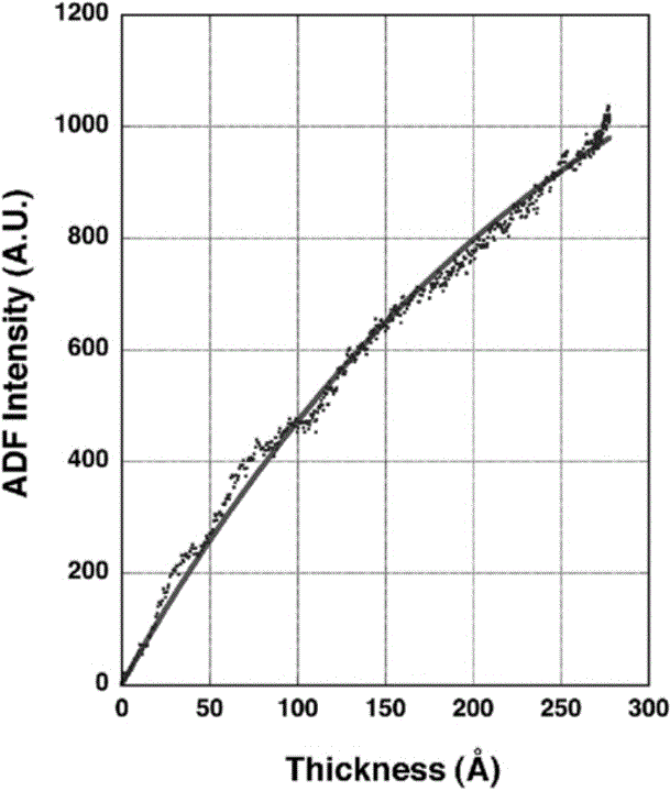
Figure 1197a shows a profile of ADF-STEM intensity obtained from GaN materials in different thickness.
From this plot the electron scattering cross-section in Equation 1197 can be
determined.

Figure 1197a. ADF-STEM intensity from GaN materials in different thickness. [9] |
Table 1197b. Dependence of intensity in annular dark-field (ADF) images.
Factors |
Dependence |
| Smaller camera length |
Lower
intensity |
| Larger effective inner detection angle |
Lower
intensity |
| Large angles, i.e. >>100 mrad |
Intensity ∝ Number of atom scattering * Z2 (Z is atomic number) [6] |
Table 1197c. Electron scattering in annular dark-field (ADF) images.
Factors |
Dependence |
| Large angles, i.e. >>100 mrad |
Rutherford scattering by
the atomic nuclei is the dominant scattering mechanism |
Table 1197d. Applications in annular dark-field (ADF) images.
Applications |
Detection angles |
Characteristics |
| HR-STEM |
Low, ~30 mrad |
Rutherford scattering and Bragg scattering |
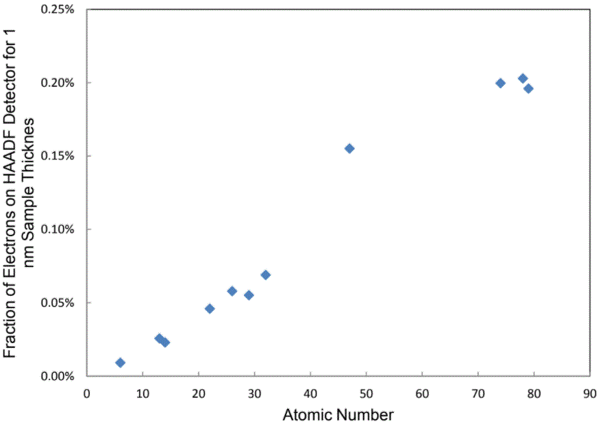
Figure 1197b shows the fraction, ε, of electrons scattered onto the HAADF-STEM detector per
nanometer of sample thickness for different pure elements. The angular range for the ADF collection is from 53 mrad to 230 mrad.

Figure 1197b. Fraction ε of electrons scattered onto the HAADF-STEM detector per
nanometer of sample thickness for different pure elements. [10] |
[1] Pennycook SJ et al. 1992 Scanning Microsc. Suppl. 6, 233.
[2] Treacy MMJ and Gibson JM 1993 Ultramicroscopy 52, 31.
[3] Cowley JM and Huang Y 1992 Ultramicroscopy 40, 171.
[4] Pennycook SJ and Boatner LA 1988 Nature 336, 565.
[5] Walther T and Humphreys CJ 1997 Inst. Phys. Conf. Ser. 153, 303.
[6] Treacy M M J, Howie A and Wilson CJ 1978 Philos. Mag. A 38, 569.
[7] Rutherford E 1911 Philos. Mag. 21, 669.
[8] R.D. Heidenreich, Fundamentals of Transmission Electron
Microscopy, Wiley, New York, 1964, p. 31.
[9] S. Bals, B. Kabius, M. Haider, V. Radmilovic, C. Kisielowski, Annular dark field imaging in a TEM, Solid State Communications 130 (2004) 675–680.
[10] Biao Yuan, Direct Measurement of Thicknesses, Volumes or Compositions of Nanomaterials by Quantitative Atomic Number Contrast in High-angle Annular Dark-field Scanning Transmission Electron Microscopy, University of Central Florida, thesis, (2012).
|