=================================================================================
It was suggested that the column diameter (from objective lens to intermediate lenses) in a (S)TEM equipped with a Cs-corrector was increased from 250 to 300 mm to enhance the column stiffness. [1]
The schematic illustration in Figure 2241 presents the column part in typical (S)TEM systems.
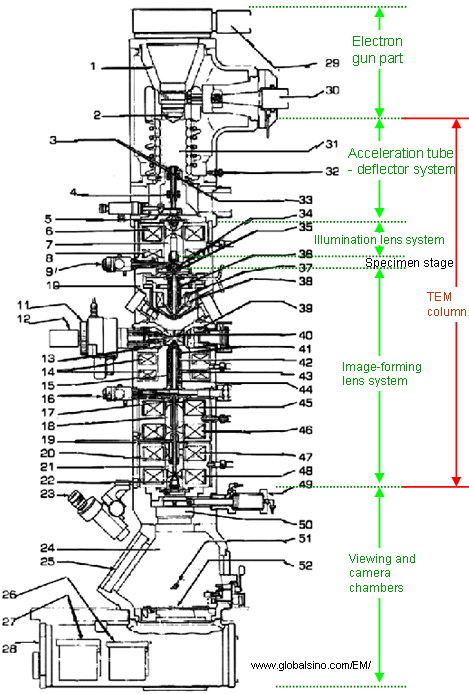 |
1. Electron gun
2. Wehnelt unit
3. Anode
4. Electron gun second beam delector coil
5. Anode chamber isolation valve
6. 1st condenser lens coil
7. Condenser polepiece
8. 3rd condenser lens coil
9. Condenser aperture assembly
10. Specimen chamber
11. Goniometer
12. specimen holder
13. Stigmator screening cylinder
14. Objective lens coil
15. Objective lens liner tube
16. Field limiting aperture
17. Intermediate lens stigmator
18. Intermediate polepiece
19. Intermediate lens linear tube
20. Projector lens beam deflector coil
21. Projector upper polepiece
22. Projector lower polepiece
23. Binoculars
24. Viewing chamber
25. Viewing window
26. Dispensing magazine
27. Receiving magazine
28. Camera chamber
29. Lift arm
30. HT cable to high voltage tank
31. Anode chamber, or called acceleration tube
32. Gas inlet
33. Electron gun 1st beam deflector coil
34. Condenser lens stigmator coil
35. Spot alignment coil
36. Condenser lens 1st beam deflector coil
37. Condenser lens 2nd beam deflector coil
38. Condenser minilens (CM) lens coil
39. Stage heater
40. Objective polepiece
41. Objective lens stigmator coil
42. 1st image shift coil
43. Objective minilens (OM) lens coil
44. 2nd image shift coil
45. 1st intermediate lens coil
46. 2nd intermediate lens coil
47. 3rd intermediate lens coil
48. Projector lens coil
49. Viewing chamber isolation valve
50. High resolution diffraction chamber
51. Small screen
52. Large screen |
Figure 2241. Schematic illustration of the structure of typical TEM systems (e.g. JEM-2010F
here). |
In order that the electron beam is not scattered significantly by the air molecules in the TEM column, the pressure in the column must be <~0.1 Pa.
[1] Takeo Sasaki, Hidetaka Sawada, Fumio Hosokawa, Yuji Kohno, Takeshi Tomita, Toshikatsu Kaneyama, Yukihito Kondo, Koji Kimoto, Yuta Sato, and Kazu Suenaga, Performance of low-voltage STEM/TEM with delta corrector and cold field emission gun, Journal of Electron Microscopy 59(Supplement): S7–S13 (2010).
|