| A TEM with a phase plate in combination with appropriate wave reconstruction method has advanced applications to structural biology [1–4]. To achieve these applications, one needs to satisfy:
i) Fabricate a reliable phase plate;
ii) The TEM functions cannot be degraded by the phase plate;
iii) The phase plate needs to be accurately (with nanometer precision) positioned at the back focal plane of the objective lens so that the electron beam can pass through the device precisely and can be phase shifted;
iv) Cryo-EM and low dose imaging conditions are needed in order to preserve the vulnerable bio-structure during imaging [5–6].
Some types of phase plate designs have been suggested:
i) Zernike-type phase plate [7],
ii) Hilbert-type phase plate [8–11].
Due to the limited space in the pole piece gap in the TEM columns, a phase plate in TEMs has traditionally required a modification of the optical design of the TEM. However, TEM phase plate loading systems, which can be installed in commercial TEMs without modification of their optical design, have recently been developed. For instance, Shiue and Hung [12] presented a system with a loading monitoring function allowing the user to quickly and safely position an electrostatic phase plate, in three-axis with <10 nm precision, from a computer. Figure 2567 shows the TEM phase plate loading system.
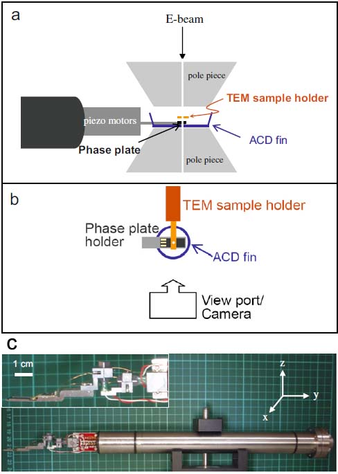
Figure 2567. (a) Schematic side-view and (b) schematic top-view of the relative positions of the components of a TEM phase plate loading system around the pole pieces. A camera can be installed on an emptied aperture port. ACD fin stands for an anti-contamination device fin. (c) The phase plate loading rod. The inset shows the magnified photograph of the three-axis nano-positioner connected with a phase plate holder. Adapted from [12]
[1] R. M. Glaeser, Cryo-electron microscopy of biological nanostructures, Physics
Today 61 (2008) 48–54.
[2] H. Stahlberg, T. Walz, Molecular electron microscopy: state of the art and
current challenges, ACS Chemical Biology 3 (2008) 268–281.
[3] R. Danev, K. Nagayama, Transmission electron microscopy with Zernike phase
plate, Ultramicroscopy 88 (2001) 243–252.
[4] Y. Fukuda, Y. Fukazawa, R. Danev, R. Shigemoto, K. Nagayama, Tuning of the
Zernike phase-plate for visualization of detailed ultra structure in complex
biological specimens, Journal of Structural Biology 168 (2009) 476–484.
[5] J. Shiue, C. -S. Chang, S. -H. Huang, C. -H. Hsu, J. -S. Tsai, W. -H. Chang, Y. -M. Wu,
Y. -C. Lin, P. -C. Kuo, Y. -S. Huang, Y. Hwu, J.-J. Kai, F. -G. Tseng, F. -R. Chen, Phase
TEM for biological imaging utilizing a Boersch electrostatic phase plate:
theory and practice, Journal of Electron Microscopy 58 (2009) 137–145.
[6] K. Nagayama, R. Danev, Phase-plate electron microscopy: a novel imaging
tool to reveal close-to-life nano-structures, Biophysical Reviews 1 (2009)
37–42.
[7] K. Nagayama, Development of phase plates for electron microscopes and their
biological application, European Biophysics Journal 37 (2008) 345–358.
[8] R. Danev, H. Okawara, N. Usuda, K. Kametani, K. Nagayama, A novel phase-contrast transmission electron microscopy producing high-contrast topographic images of weak objects, Journal of Biological Physics 28 (2002)
627–635.
[9] Y. Kaneko, R. Danev, K. Nitta, K. Nagayama, In vivo subcellular ultrastructures
recognized with Hilbert differential contrast transmission electron microscopy, Journal of Electron Microscopy 54 (2005) 79–84.
[10] R. Danev, K. Nagayama, Complex observation in electron microscopy: IV.
Reconstruction of complex object wave from conventional and half plane
phase plate image pair, Journal of the Physical Society of Japan 73 (2004)
2718–2724.
[11] B. Barton, F. Joos, P. R. Schroder, Improved specimen reconstruction by Hilbert
phase contrast tomography, Journal of Structural Biology 164 (2008)
210–220.
[12] Jessie Shiue, Shao-Kang Hung, A TEM phase plate loading system with loading monitoring and nano-positioning functions, Ultramicroscopy 110 (2010) 1238–1242.
|
