|
This book (Practical Electron Microscopy and Database) is a reference for TEM and SEM students, operators, engineers, technicians, managers, and researchers.
|
=================================================================================
The sigma (σ) bonds are formed by axial overlap of half-filled atomic orbitals of atoms taking part in bonding. Sigma bonds are stronger than Pi bonds. In chemistry, sigma bonds (σ bonds) are actually the strongest type of covalent chemical bonds.
The configuration of the valence electrons in carbon is a combination (called hybrids) of s and p orbitals. Graphite is one example with an sp2 type, while diamond is of an sp3 form. Figure 2626a illustrates the hybridization process for diamond. During this process, the energy levels of the 2s are raised while the 2p levels are reduced, resulting in a more favourable lower energy system. In this case, these four valence electrons form σ bonds.
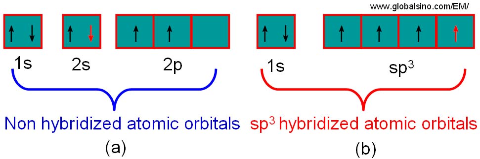
Figure 2626a. Schematic illustration of the process of hybridisation for carbon. Each carbon atom in diamond forms a series of sp3 hybridised atomic orbitals.
The sp2 bonded solids have both σ/σ* and π/π* states available to the electrons, while for sp3 bonded solids only the σ/ σ* states present. For the diamond and graphite, electron transitions to these states generate many of the characteristic features in EEL spectra. For instance, in core loss spectra from diamond, the excitation of the 1s electrons to the σ* states generates the carbon k edge peak. For graphite, the transitions of the inner shell electrons into unoccupied π* states give a peak prior to the edge onset. In the valence band, valence electron transitions into the π* states also produce a peak at around 6 eV. Figure 2626b shows carbon (C) K edge from materials containing the carbonate anion (calcite, siderite, desautelsite, and hydrotalcite) and three allotropes of C (amorphous , graphite, and diamond). EELS profiles of amorphous C and graphite present basically the same edge shapes, with the first peak at 285 eV induced by transitions to the π* molecular orbital due to the presence of sp2 bonding, and the second, more intense peak at 290 eV induced by transitions to σ* orbitals. In diamond the bonding between the C atoms can be interpreted in terms of tetrahedrally directed sp3 hybrid orbitals, and the first peak with a maximum at 292.6 eV is identified as arising from transitions to molecular orbitals of σ* character.
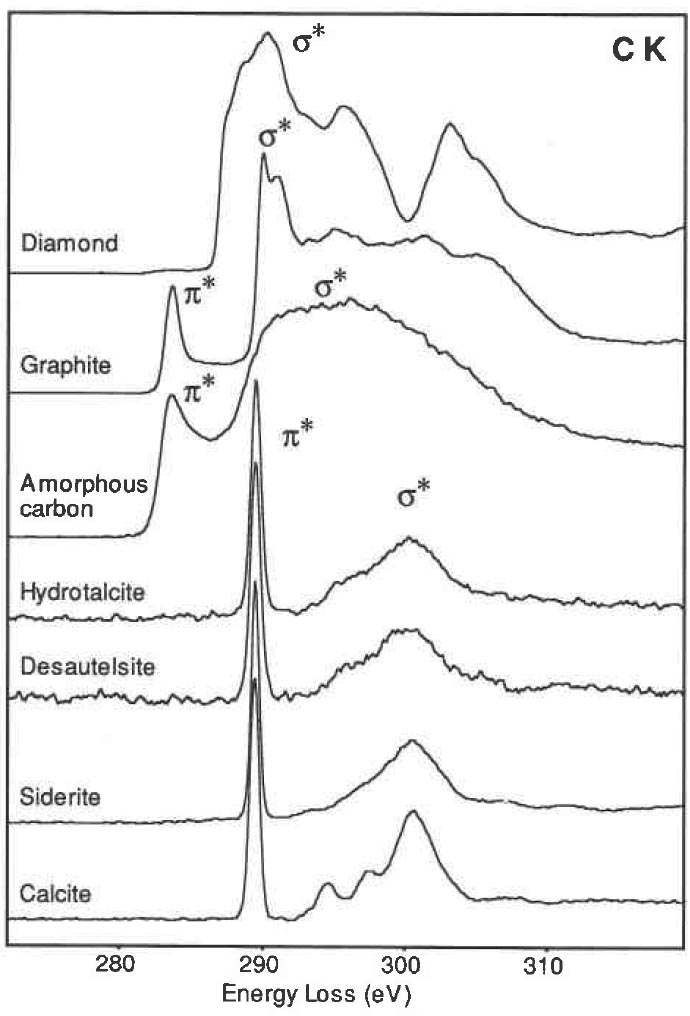
Figure 2626b. Carbon K edge from calcite, siderite, desautelsite, hydrotalcite, amorphous, graphite, and diamond.
|