=================================================================================
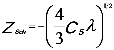
Practically, the large, and positive third-order spherical aberration can be combined with a optimized defocus setting (namely, sample height Z = C1,0) to form the Scherzer λ/4 phase plate,
 ---------------------------- [2769a] ---------------------------- [2769a]
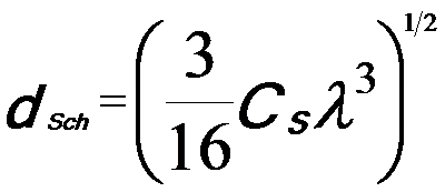
So that the point resolution is,
 ---------------------------- [2769b] ---------------------------- [2769b]
In this case, the object information up to a spatial frequency 1/dSch is transferred with the same sign of the aberration function, yielding dark-atom contrast for a TEM specimen that can satisfy weak phase object approximation (WPOA) condition.
Based on Cs correction, extending Scherzer’s point resolution to information limit of the microscope can optimize the positive phase contrast from a weak-phase object. Assuming gSch = gmax and a defocus of ZSch = -8/(3λg2max)
, one can obtain, [1]
 ---------------------- [2769c] ---------------------- [2769c]
The resulting contrast delocalization is given by,
 ---------------------- [2769d] ---------------------- [2769d]
In this case, the atoms are also observed in dark contrast with respect to the background.
In summary, due to the positive spherical aberration in traditional TEMs, the only way to produce phase contrast from a thin object is to use an underfocus setting. The resulting aberration function from underfocus is negative and the phase change is positive. The positive phase contrast from a weak phase object is thus dark with respect to the background (mean intensity).
The phase-contrast term -isin2πχ(g) in the contrast transfer function becomes zero if CS=0 and Δf=0, while the amplitude-contrast term cos2πχ(g) is maximum, 1. Therefore, for aberration-corrected microscope, the TEM images present atomic structures by amplitude-contrast rather than by phase-contrast, and thus in HRTEM images the projected atom column are imaged in bright contrast on a dark background.
Table 2769. More readings.
| TEM contrast and underfocus/defocus |
page4214 |
| Contrast reversal of bright field TEM/STEM images |
page2881 |
[1] Lentzen, M., Jahnen, B., Jia, C.L., Thust, A., Tillmann, K. &
Urban, K. (2002). High-resolution imaging with an aberrationcorrected
transmission electron microscope. Ultramicroscopy
92, 233–242.
|
 ---------------------------- [2769a]
  ---------------------------- [2769b]