|
This book (Practical Electron Microscopy and Database) is a reference for TEM and SEM students, operators, engineers, technicians, managers, and researchers.
|
=================================================================================
Figure 2935 shows the contrast of high resolution HAADF STEM image, as a function of crystal tilt from 0° to 4° (0-67 mrad), taken from a (100) SrTiO3 crystal in Cs-corrected STEM mode. A small crystal tilt (e.g. 0-15 mrad) can reduce the contrast of high resolution ADF images by a factor of 2 because of dynamical scattering. Moreover, this effect also depends on the sample thickness, which was 15 nm here.
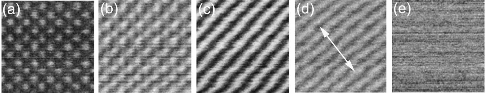
Figure 2935. STEM-ADF image of (100) SrTiO3 as a function of crystal tilt. (a) 0°; (b) 1°; (c) 2°; (d) 3°; and (e) 4°. The tilt axis as marked in (d) is near the [110]. Adapted from [1]
[1] H. Inada, D.Su, R. F. Egerton, M.Konno, L.Wu, J.Ciston, J.Wall, Y.Zhu, Atomic imaging using secondary electrons in a scanning transmission electron microscope: Experimental observations and possible mechanisms, Ultramicroscopy 111(2011)865–876.
|