Back Focal Plane of Projector Lens in TEM - Practical Electron Microscopy and Database - - An Online Book - |
||||||||
| Microanalysis | EM Book http://www.globalsino.com/EM/ | ||||||||
| This book (Practical Electron Microscopy and Database) is a reference for TEM and SEM students, operators, engineers, technicians, managers, and researchers. | ||||||||
| ================================================================================= | ||||||||
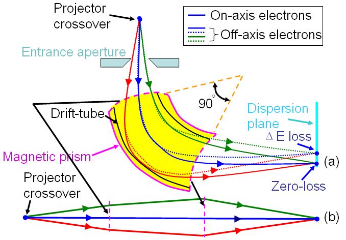
In Figure 3162 (a), the spectrum is formed in the dispersion plane, consisting of a distribution of electron counts (I) versus energy loss (ΔE). All the electrons suffering the same energy loss but traveling in both on-axis and off-axis directions are directed to a focus in the dispersion plane of the spectrometer, which acts as a homogenous magnetic lens as shown in the equivalent schematics in Figure 3162 (b). The object plane of the spectrometer is typically set at the back focal plane (crossover) of the projector lens.
Figure 3162. (a) Schematic showing magnetic prism, and (b) Equivalent schematics of the magnetic prism. Electrons at various kinetic energies (due to energy losses induced by interaction with TEM specimen) are focused at the energy-dispersive plane of the spectrometer.
|
||||||||
| ================================================================================= | ||||||||
| The book author (Yougui Liao) welcomes your comments, suggestions, and corrections, please click here for submission. If you let book author know once you have cited this book, the brief information of your publication will appear on the “Times Cited” page. | ||||||||
|
|
||||||||