=================================================================================
Inelastic scattering of incident electrons occurring in materials is physically delocalized within a certain lateral distance. This phenomenon fundamentally induces a physical limit on the spatial resolution of the inelastic signal, even though the signal is recorded with an ideal instrument.
In EFTEM imaging, an important contribution for low-loss imaging is the delocalization of the inelastic scattering process itself, while at much higher energy-losses the resolution-limiting parameter is usually the chromatic aberration. For instance, to lower the effect of the chromatic aberration, the accelerating voltage needs to be 300 kV if one expects a resolution better than 0.3 nm for >100 eV energy-loss [1].
The inelastic delocalization factor R can be estimated by the root mean square impact parameter drms proposed by Pennycook [2],
 --------------------------------------------[3409] --------------------------------------------[3409]
where,
drms -- The root mean square impact parameter.
h -- Planck-constant
ν -- Wave-frequency
ΔE -- Energy loss
λ -- wavelength of incident electron beam
θo -- Semiangle
due to the objective aperture size, namely objective lens collection semi-angle
θE -- The characteristic scattering semiangle.
The scattering localization in the excitations of inner-shell electrons is much better than those in the direct excitations of valence electrons. In the interaction of incident electrons with solids, valence electrons are delocalized and produce collective excitations called plasmons. Based on a non-relativistic theory, Allen and Rossouw [3] calculated the inelastic object functions for K-shell ionization. The calculated object functions for the K-shell excitations of some elements such as Te, Cd, As and Ga indicate that the localization of the object is less than 1 Å [4].
Figure 3409 shows the length of the delocalization of inelastic scattering as a function of the primary electron beam energy, normalized to E0 = 200 keV.
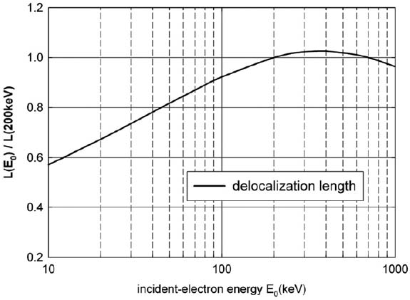
Figure 3409. Delocalization length as a function of primary electron beam energy.
[5]
[1] R.F. Egerton, Electron Energy-Loss Spectroscopy in the Electron Microscope, 2nd Edition, Plenum Press, New York, 1996.
[2] Pennycook S. J., 1982. High resolution electron microscopy and
microanalysis. Contemp. Phys., 23, 371–400.
[3] L.J. Allen, C.J. Rossouw, Phys. Rev. B 42 (1990) 11644–11654.
[4] N.D. Browning, S.J. Pennycook, Microbeam Anal. 2 (1993) 81–89.
[5] H. Inada, D.Su, R. F. Egerton, M.Konno, L.Wu, J.Ciston, J.Wall, Y.Zhu, Atomic imaging using secondary electrons in a scanning transmission electron microscope: Experimental observations and possible mechanisms, Ultramicroscopy 111(2011)865–876.
|