=================================================================================
One type of TEM tomography methods is called confocal electron microscopy. The main mechanism of this technique is that the STEM probes (especially Cs-corrected probes) have a short depth of focus. This method constrains the depth of field/focus in the image to a very thin plane using a confocal aperture before the image plane. A set of images are recorded over a range of defocus. This method is also called depth sectioning achieved
by a focal series [1]. In this case, it is possible to obtain detailed structural information from within the crystal volume.
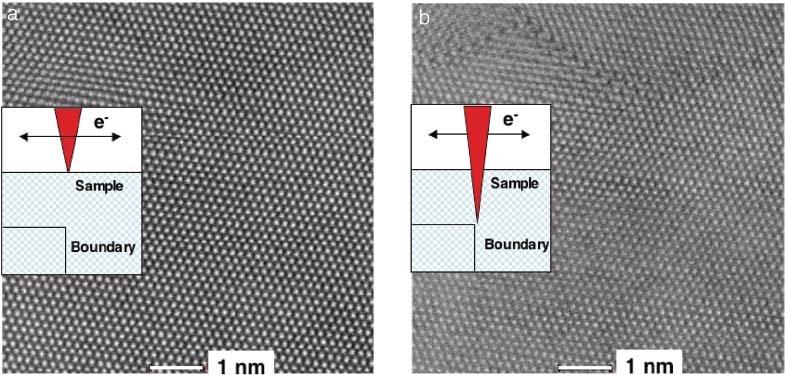
As an example, Figure 3981 shows HAADF STEM images of a gold [111] crystal with an electron beam focused on the crystal surface and defocused at 6 nm. The extended crystal defect, which is a buried Σ3{112} grain boundary, is visible only when the beam is focused into the crystal at a defocus of 6 nm.

Figure 3981. HAADF STEM images of gold [111]: (a) STEM probe focused on sample surface (indicated by the inset); (b) Grain boundary in the lower section of the foil is visible only when the probe is focused 6 nm into the sample.
Those images were taken in a microscope with an aberration corrector. [2]
[1] Borisevich, A.Y., Lupini, A.R., Travaglini, S. & Pennycook, S.J. (2006). Depth sectioning of aligned crystals with the aberrationcorrected
scanning transmission electron microscope. J Electron
Microsc 55, 7–12.
[2] C. Kisielowski, B. Freitag, M. Bischoff, H. van Lin, S. Lazar, G. Knippels, P. Tiemeijer, M. van der Stam, S. von Harrach, M. Stekelenburg, M. Haider, S. Uhlemann, H. Müller, P. Hartel, B. Kabius, D. Miller, I. Petrov, E. A. Olson, T. Donchev, E.A. Kenik, A. R. Lupini, J. Bentley, S.J. Pennycook, I. M. Anderson, A.M. Minor, A.K. Schmid, T. Duden, V. Radmilovic, Q. M. Ramasse, M. Watanabe, R. Erni, E.A. Stach, P. Denes, and U. Dahmen, Detection of Single Atoms and Buried Defects in Three Dimensions by Aberration-Corrected Electron Microscope with 0.5-Å Information Limit, Microsc. Microanal. 14, 469–477, 2008.
|
