=================================================================================
The extent of information transfer and the spatial resolution can be calculated by frequency space obtained from Young’s fringes produced by an image shift during imaging exposure. Figure 4150 shows the Fourier transforms of HRTEM (high-resolution TEM) images of gold (Au) nanoparticles on an amorphous carbon film. A monochromator was installed in the TEM system. When the monochromator was turned off, the Young’s fringes extended only to about 70 pm. When the monochromator was turned on and thus the effect of chromatic aberration was reduced, the Young’s fringes extended beyond 50 pm (red circle) in all directions, reflecting that this microscope with the monochromator has 50 pm spatial resolution.
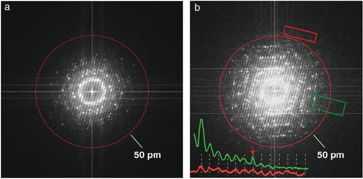
Figure 4150. HRTEM performance demonstrated by Young’s fringe experiments with gold nanoparticles on a carbon film: (a) With monochromator turned off, the Young’s fringes extended to about 70 pm; (b) With monochromator turned on, the Young’s fringes extended beyond 50 pm. The inset line traces are obtained from the areas outlined in green and red boxes [1].
[1] C. Kisielowski, B. Freitag, M. Bischoff, H. van Lin, S. Lazar, G. Knippels, P. Tiemeijer, M. van der Stam, S. von Harrach, M. Stekelenburg, M. Haider, S. Uhlemann, H. Müller, P. Hartel, B. Kabius, D. Miller, I. Petrov, E. A. Olson, T. Donchev, E.A. Kenik, A. R. Lupini, J. Bentley, S.J. Pennycook, I. M. Anderson, A.M. Minor, A.K. Schmid, T. Duden, V. Radmilovic, Q. M. Ramasse, M. Watanabe, R. Erni, E.A. Stach, P. Denes, and U. Dahmen, Detection of Single Atoms and Buried Defects in Three Dimensions by Aberration-Corrected Electron Microscope with 0.5-Å Information Limit, Microsc. Microanal. 14, 469–477, 2008.
|