=================================================================================
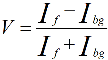
The concept of contrast is defined as the relative differences in observed intensities in an object, representing visual discrimination of the object. For any type of signals (e.g. TEM, SEM and STEM images, EDS, and EFTEM) from a feature within an EM specimen, in order to detect the feature in an image the contrast needs to be 3 ~ 5 times greater than the inherent noise signal. The image contrast, sometimes called visibility (V), is given in terms of the maximum and minimum intensities as,
 ----------------------------------------- [4297a] ----------------------------------------- [4297a]
Moreover, the detection and visualization of a feature depend on both the contrast and the resolution.
The visibility of a specific feature can be quantified by,
 ----------------------------------------- [4297b] ----------------------------------------- [4297b]
where,
Ic and Ibg -- The pixel intensities of the feature and of the
background, respectively.
For HRTEM imaging, CTF (contrast transfer function) partially changes the contrast of the images by adjusting Scherzer defocus. Assuming Cs and λ are constant for a specific microscope, Figure 4297 shows CFT T(|g|) = sin(χ) at n Scherzer defoci, given by,
 ------------------------------- [4297c]
where,
n -- 0, 0.5, 1, 1.2 (extended), 2, and 10
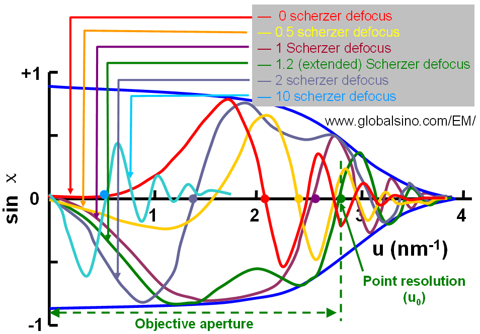
Figure 4297. Contrast transfer function (CFT) at 0, 0.5, 1, 1.2 (extended), 2, and 10 Scherzer defoci.
For instance, 0.5 Scherzer defocus is a defocus condition given minimum contrast and 2 scherzer defocus is the second-passband defocus, giving positive CTF to produce a negative phase contrast (atoms are in white contrast).
|