Descan of Electron Beam in (S)TEM - Practical Electron Microscopy and Database - - An Online Book - |
||||||||
| Microanalysis | EM Book https://www.globalsino.com/EM/ | ||||||||
| ================================================================================= | ||||||||
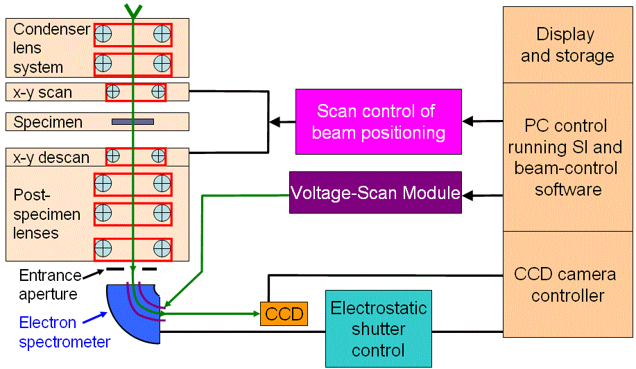
Figure 1765 shows the schematic illustration of a STEM spectrum imaging system. The focused electron probe is scanned across the specimen and a couple of post-specimen coils is used to descan.
In the technique of precession electron diffraction (PED), to obtain a stationary diffraction pattern instead of ring reflections, a simultaneous descan of the diffracted beam is also needed by means of image shift TEM coils.
|
||||||||
| ================================================================================= | ||||||||
|
|
||||||||