Table 3110. Comparison between EDS and WDS techniques.
|
EDS |
WDS |
Full name |
Eenergy dispersive X-ray spectroscopy |
Wavelength dispersive X-ray spectroscopy |
Principle |
Measures the energy of X-ray photon |
Determine the wavelength of X-ray |
Spectrum range |
Measures across a broad range of energies in the spectrum |
Measures specific regions within the spectrum |
Efficiency |
More efficient for an unknown specimen |
More efficient for the concentration of specific, known element |
Speed |
Quicker for large energy ranges |
Slower for large energy ranges |
Throughput of X-rays |
Lower |
Higher |
Spatial resolution |
The same |
|
Energy resolution |
Lower: ~ 125 eV |
Higher: ~5-10 eV |
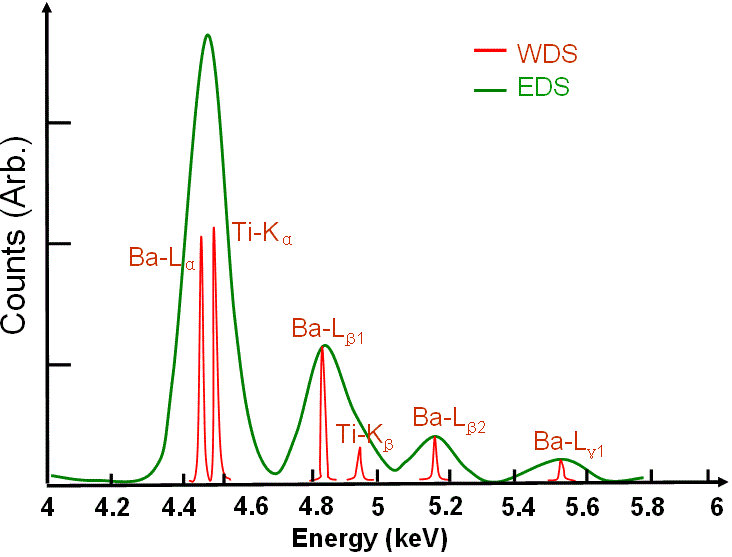
EDS and WDS profiles of BaTiO3. The green spectrum presents a standard EDS of of BaTiO3, while the red one presents a standard WDS.
EDS shows the overlapped Ba Lα-Ti Kα and Ba Lβ1-Ti Kβ peaks. |
Sensitivity |
OK |
Better due to separation of overlapping peaks |
Peak-to-background ratio |
Lower |
Higher |
Specimen limitation |
Gases cannot be analyzed and liquids are limited to those that have very limited volatility and will not contaminate the column and specimen chamber because specimens must be exposed to vacuum conditions. |
Hardware |
Attached to electron microscopes |