=================================================================================
Figure 3647a shows the simulated intensity distribution patterns of 200 keV-electron probes at 100 nm of B2 (second-order axial coma), 100 nm of A2 (three-fold axial astigmatism), 1 µm of S3 (axial star aberration of the 3rd order), and 2 µm of A3 (four-fold axial astigmatism).
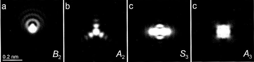
Figure 3647a. Simulated intensity distribution patterns of 200 keV-electron probes: (a) B2 = 100 nm, (b) A2 = 100 nm, (c) S3 = 1 µm, and (d) A3 = 2 µm. In this simulation, the defocus C1 was set to -3 nm, the imaginary parts of the aberrations to zero, and the illumination semi-angle to 30 mrad. [1]
Figure 3647b shows the schematic comparison of Zemlin (diffractogram)-tableau characteristics for the axial aberrations up to third orders. First-order aberration (e.g. defocus and twofold astigmatism, A1) shows the elliptical distortion even without electron beam tilting because the impact of first-order aberrations does not depend on the tilt angle. For the aberrations of higher orders (n≥2), such as second-order axial coma B2, three-fold astigmatism A2, third-order spherical aberration C3 (>0), third-order star aberration S3 and four-fold astigmatism A3, there is no elliptical distortion observable for the un-tilted case. In these cases, only at illumination tilts the characteristic distortion due to the aberrations becomes discernible. Note that the higher-order aberrations have equal symmetries to the ones in Figure 3647a as discussed in page3740.
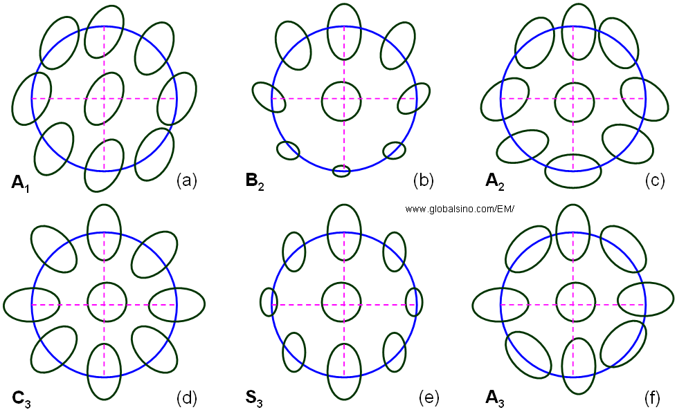
Figure 3647b. Schematic representation of Zemlin (diffractogram)-tableau characteristics for the axial aberrations up to third order.
After the corrections of lower order aberrations, the corrector normally introduces or leaves high order aberrations. For instance, the Ronchigram with a 54 mR aperture indicated in Figure 3647c shows the left 4th order coefficient of order 0.5 - 2.0 mm limiting the probe shape and size after 3rd order aberration correction by Nion quadrupole-octupole corrector. The pattern is strongly 4-fold symmetry with weak 2-fold structure in the opposing corners. Note that this uniform 4-fold symmetry was actually imposed by the octupoles in the corrector, so called parasitic aberrations.
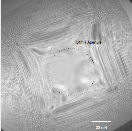
Figure 3647c. Ronchigram obtained with 4th order coefficients corrected to < 60 µm [2].
Note that an octupole always generates parasitic aberrations because it has cubic radial dependence together with four-fold azimuthal symmetry (four-fold astigmatism).
[1] Simulation of Rolf Erni.
[2] P. E. Batson, Control of Parasitic Aberrations in Multipole Corrector Optics, Microsc Microanal 14(Suppl 2), 2008.
|