=================================================================================
The condenser lens system converges the electron beam to a focus at or near the plane of the specimen. In the arrangement of the basic imaging systems of conventional transmission electron microscopy (CTEM), an electron beam emitted by a source radiates the TEM specimen, usually through an illuminating system of lenses as shown in blue in Figure 3763. The incident electrons interact with the specimen and thus are scattered. The electron beam after interaction is then focused by the objective lens, forming an image. The image is further magnified by projector lenses to produce an image at proper magnifications. Note that in modern TEM systems additional lenses, intermediate lenses are also placed between objective lens and projector lenses.
For scanning transmission electron microscopy (STEM), the same diagram can be used with a reversed direction of the electron beam also shown in Figure 3763. The electron source for STEM replaces the TEM detection system (image plane). In modern STEM systems, a field-emission gun can be used to produce an electron source as small as 10 nm in diameter. One or more projector lenses in TEM (acting as condenser lenses in STEM) with a short focal length demagnify the source to provide the flexibility in the beam parameter. The beam is then demagnified further by the main lens in the systems, which is objective lens to form a small probe of 0.2 – 1 nm on the specimen. Therefore, a STEM probe can be considered as a demagnified image of the source. Note that the focal length of the objective lens can be as small as 1 mm. The scanning coils are deflector coils, built into the bore of the objective lens, and serve to scan the electron probe over the specimen. The detector on the STEM image plane collects the electrons transmitted, or scattered, by the specimen.
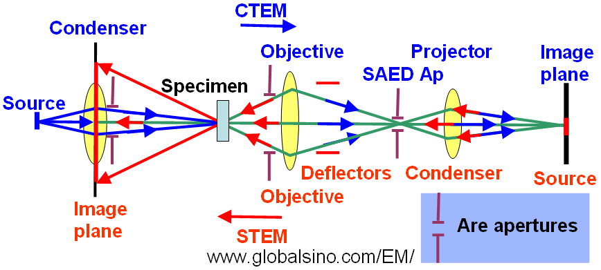
Figure 3763. Schematic illustration of imaging geometries of TEM and STEM systems. The green lines show the common paths of the rays of CTEM and STEM illuminations. The blue arrows indicate the rays for the TEM illumination, while the red arrows indicate the directions of STEM rays. The STEM image is formed on the left-hand side, while the CTEM image is on the right-hand side. The yellow lenses are the common lenses for both STEM and CTEM systems.
The condenser lens system provides variable probe-convergence angles in STEM mode and adjustable parallel illumination in TEM mode. Note that the commercially standard condenser lens system in FEI TEMs has three condenser lenses.
Astigmatism in condenser lens is important because it reduces the coherence of the electron beam, while astigmatism in objective lens is important because it induces a serious degradation of spatial resolution.
There are mainly two steps to correct the astigmatisms in TEMs:
i) Correct the astigmatisms of the diffraction (inter-mediate, IL) and condenser (CL) lenses. Before the astigmatism correction for objective lens (OL) , the IL and CL astigmatisms should be corrected. The CL astigmatism must especially be corrected (without objective aperture) to ensure the proper correction of the OL astigmatism.
ii) Correct the OL astigmatism.
In STEM mode, the probe size, or called beam size and probe diameter, of modern microscopes can be defined as the full-width-at-half-maximum (FWHM) which contains 50-75% of the beam current. In this case, the probe is first focused at the eucentric height, and then focused on the viewing screen or CCD camera using the second condenser lens, C2.
From the EM operation point of view, the probe size, d, can only be reduced by using a smaller condenser aperture or by increasing the excitation of C1 lens.
|