|
|
Spatial Resolutions of Various Techniques
- Practical Electron Microscopy and Database -
- An Online Book -
|
|
http://www.globalsino.com/EM/
|
|
This book (Practical Electron Microscopy and Database) is a reference for TEM and SEM students, operators, engineers, technicians, managers, and researchers.
|
=================================================================================
Figure 3929. Comparison of spatial resolutions between different techniques.
| Probe |
Signal |
Technique |
Best Spatial Resolution* (nm) |
| Light |
Light |
Bare human eyes |
10, 000 |
| Electron |
Electron |
Scanning electron microscope (SEM) |
< 10 |
| Electron |
Electron |
Selected-area electron diffraction (SAED) |
10 - 1000 |
| Electron |
Electron |
Convergent beam electron diffraction (CBED) |
10 - 1000 |
| Electron |
Electron |
Auger electron spectroscopy (AES) |
< 2 |
| Electron |
Electron |
Transmission electron microscope (TEM) |
< 0.1 |
| Electron |
Electron |
Scanning TEM (STEM) |
< 0.1 |
| Electron |
Electron |
Electron energy-loss spectroscopy (EELS) |
< 1 |
| Electron |
Photon |
Cathodoluminescence (CL) |
|
| Electron |
Photon |
Eenergy Dispersive X-ray Spectra ( EDS) on TEM |
2 - 20 |
| Electron |
Photon |
Eenergy Dispersive X-ray Spectra ( EDS) on SEM |
1, 000 |
| Electron |
Photon |
X-ray emission spectroscopy (XES) |
1.5 – 10 |
| Ion |
Ion |
Secondary ion mass spectrometry (SIMS) |
< 50 |
| Ion |
Ion |
Local electrode atom probe (LEAP) |
< 0.1 |
| Ion |
Ion |
Rutherford backscattering spectroscopy (RBS) |
~ 1, 000 |
| Ion |
Photon |
Proton-induced x-ray emission (PIXE) |
< 500 |
| Photon |
Photon |
X-ray absorption spectroscopy (XAS) |
< 20 |
| Photon |
Photon |
X-ray diffraction (XRD) |
< 25 |
| Photon |
Photon |
X-ray fluorescence spectroscopy (XRF) |
|
| Photon |
Electron |
Ultraviolet photoelectron spectroscopy (UPS) |
< 1, 000 |
| Photon |
Electron |
X-ray photoelectron spectroscopy (XPS) |
5 – 10 |
| Photon |
Electron |
Photoelectron microscopy (PEM or PEEM) |
< 0.5 |
| Photon |
Ion |
Laser microprobe mass analysis (LAMMA) |
1, 000 |
* The best spatial resolution is the spatial resolution limit which can be reached by the modern instruments, but it is probably not the resolution limit of your instrument.
Figures 3928a and 3928b shows the typical sizes of various material defects and the capability of analytical techniques. The lowest levels of the techniques represent their spatial resolutions.
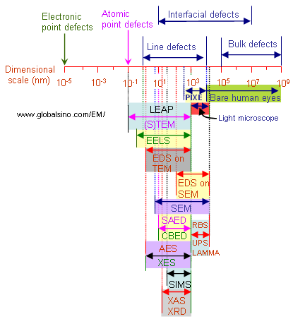
Figure 3928a. Typical sizes of various material defects and capability of analytical techniques.
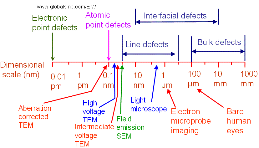
Figure 3928b. Typical sizes of various material defects and capability of main EM-related analytical techniques.
|
=================================================================================
The book author (Yougui Liao) welcomes your comments, suggestions, and corrections, please click here for submission. If you let book author know once you have cited this book, the brief information of your publication will appear on the “Times Cited” page.
|
|