Chapter/Index: Introduction | A | B | C | D | E | F | G | H | I | J | K | L | M | N | O | P | Q | R | S | T | U | V | W | X | Y | Z | Appendix
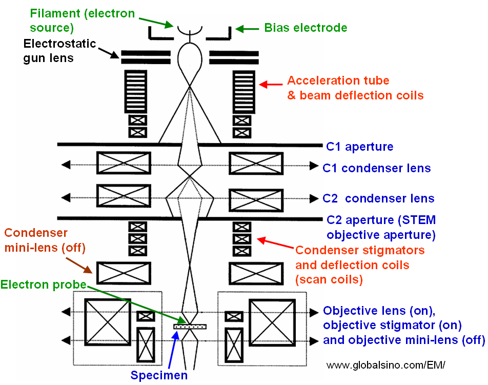
| The objective aperture itself can introduce astigmatism if it is not perfect centered on the optic axis. On the other hand, astigmatism changes from location to location in the vicinity of specimen support holder. Spatial resolution in EMs (electron microscopes) is very sensitive to objective astigmatism. Therefore, in order to obtain the best spatial resolution in EMs (especially in TEM) it is frequently necessary to carefully correct for astigmatism on the specimen. In TEM imaging, objective astigmatism is conveniently adjusted by viewing the Fresnel fringes near the edge of a small hole under overfocus condition. There are also other techniques that can be used to correct the objective astigmatism. Figure 1965a shows the structure of the electron probe-forming system in STEM mode in JEOL JEM-2010F TEMs.
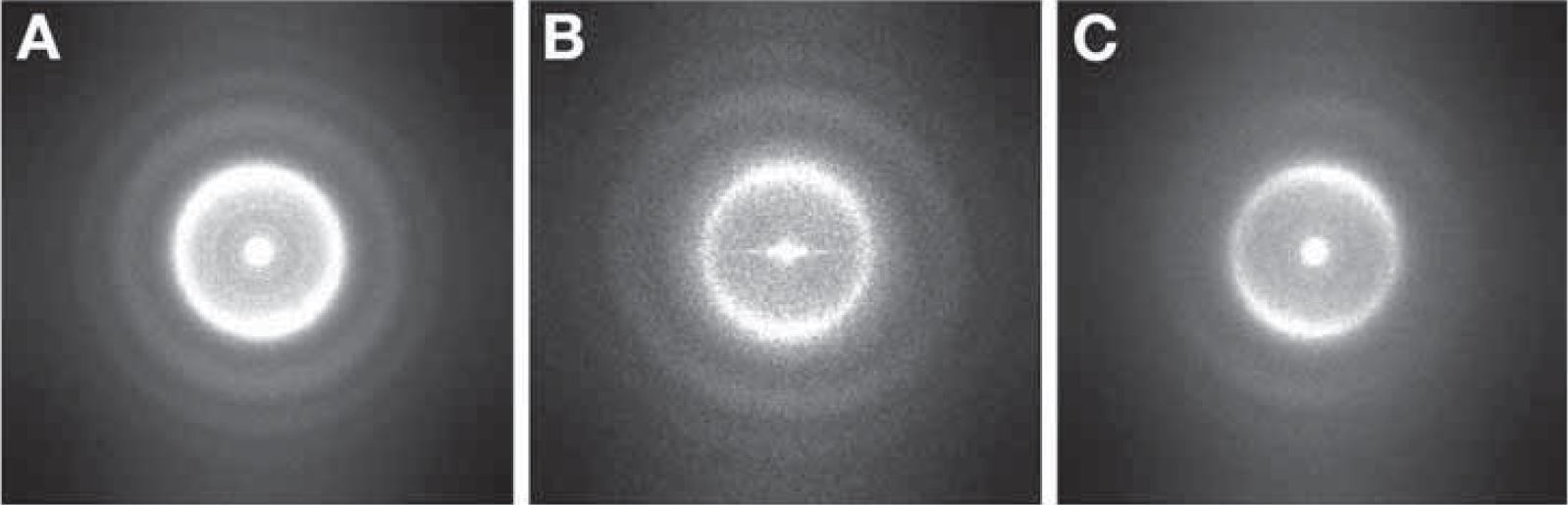
Amorphous regions in the specimen can be used to correct objective lens astigmatism. For effective correction, however, the magnification must be at least 100 k×. Figure 1965b shows Thon rings with different image qualities.
[1] Electron Microscopy: Methods and Protocols, Second Edition, 2007, Edited by John Kuo.
|