=================================================================================
The depth of focus in an electron microscope (EM) is the extent of the region around the image plane in which the image appears to be clear and sharp.
In general, the EM images will only be obviously blurry if the maximum ‘point displacement’ Δs, formed by different rays, is comparable to or greater that the spatial resolution d of an in-focus image (e.g. s in TEM and in SEM). As shown in Figure 1987, on an out-of-focus plane at height h below (or above) the in-focus plane, each point in the image will become a confused disk with a radius of s = h tanβ. Therefore, Δs can be given by,
Δs = s/M = (h/M) tan β ------------------------- [1987a]
where,
M -- The combined magnification of the entire imaging system.
β -- The convergence semi-angle, corresponding to the scattering semi-angle α.
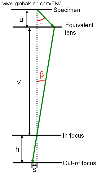
Figure 1987. Principle of depth of focus in a microscope.
This confusion leads to image blurring on the recording film or CCD. By assuming Δs = d, the depth of focus (h) of the image can then be given by,
h = Δs·M/tan β ------------------------- [1987b]
= d·M/tan β ------------------------- [1987c]
where,
d -- The spatial resolution of the EM system.
Comparing optical microscopes, one of the great advantages of SEMs is their great depth of focus because of the geometry of the beam optics.
As an example of STEM systems, Hitachi HD 2700C has a depth of focus of ~ 5 nm for HAADF mode at a probe size of 1 Å [1].
Note that spherical-aberration (Cs) correction results in a reduced depth of focus with the increase in the convergence angle θ. In general, the depth of focus can be given by,
 --------------------------------------- [1987d] --------------------------------------- [1987d]
where,
λ -- The wavelength of incident electron beam.
However, the achievable DoF in real systems depend on many sample-related factors, for instance, channelling effect.
[1] H. Inada, D.Su, R. F. Egerton, M.Konno, L.Wu, J.Ciston, J.Wall, Y.Zhu, Atomic imaging using secondary electrons in a scanning transmission electron microscope: Experimental observations and possible mechanisms, Ultramicroscopy 111(2011)865–876.
|