=================================================================================
For the In-Situ TEM/STEM method with a windowed cell, the sample is sandwiched between two electron transparent thin films (e.g. amorphous carbon films) in specially designed environmental cell (E-cell) holders. This method was first employed by Marton in 1935. [2] The gas or liquid is introduced through fine tubes running inside the sample holder rod. The phase-temperature phase diagram for water suggests that true “wet” conditions only exist at pressures of ≥ 600 Pa at 0 °C. In the range 650 to 1300 Pa, the specimen may be observed at equilibrium with water.
Figure 2569 (a) and (b) shows the schematic illustration of a fluid stage chip assembly used in TEM and STEM analysis, and Figure 2569 (c) shows a Hummingbird Scientific in-situ fluid stage single-tilt holder. The spacers prevent stiction of the silicon nitride (SiNx) membranes.
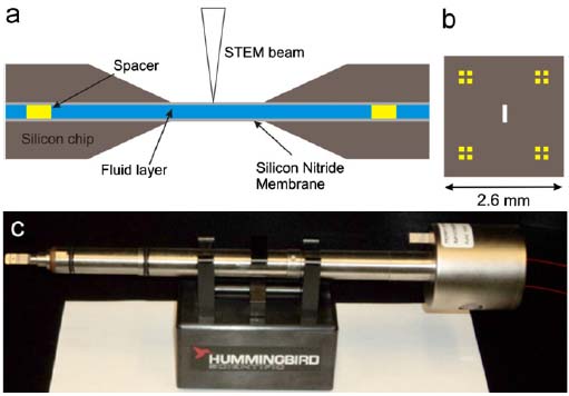
Figure 2569. (a) Schematic illustration of silicon (Si) chips used to contain a fluid layer, (b) Top-down schematic illustration of a single Si chip with gold platform spacers on each corner. (c) Hummingbird Scientific in situ fluid stage single-tilt holder. Adapted from [1]
[1] Taylor J. Woehl, Katherine L. Jungjohann, James E. Evans, Ilke Arslan, William D. Ristenpart, Nigel D. Browning, Experimental procedures to mitigate electron beam induced artifacts during in situ fluid imaging of nanomaterials, Ultramicroscopy 127 (2013) 53–63.
[2] L. Marton, Nature 133, 911 (1935).
|