|
This book (Practical Electron Microscopy and Database) is a reference for TEM and SEM students, operators, engineers, technicians, managers, and researchers.
|
=================================================================================
Chromatic aberration is a failure to bring light of different colors (wavelengths) to a common focus as shown in Figure 3641a (a). Achromatic lenses, or called achromats, are lenses that help to correct the misalignment of different color light when or after it is refracted through a lens or prism. Since different color light refracts at different angles, an achromatic lens or additional elements, for instance the flint in Figure 3641a (a), is made of different types of glass with various indices of refraction. This glass can be a weak negative glass to compensate for the dispersion of the positive crown glass in the “normal” lens as shown in Figure 3641a (b). As a result, an improved color alignment is achieved although not as good as is achieved with plan or semi-plan lens. In the field of light optics, by the middle of the 19th century, it had been widely accepted that achromatic lenses, especially achromatic objective lens, were crucial in the furtherance of serious microscopical observations. In the field of electron optics, the achromatic lenses also have been employed in some advanced electron microscopes (EMs).
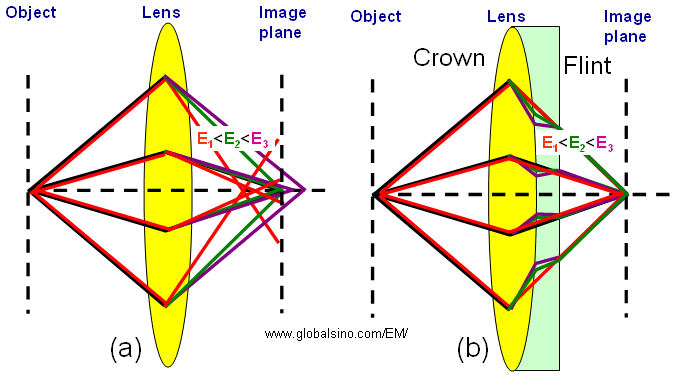
Figure 3641a. Schematic illustration of the behaviors of chromatic (a) and achromatic (b) lenses.
In advanced STEM-EELS systems, the monochromators induce an energy spectrum of the electron source at a dispersion plane, where a small fraction of the electron distribution is selected by a narrow energy-selecting slit for the measurements as shown in Figure 3641b. A drawback of this method is that the electron beam width is increased in the dispersion direction as shown in Figure 3641b (a). In some high-dispersion designs [1], this large dispersion is compensated and the electron beam is reduced to its original size by another monochromator which produces an achromatic image of the electron source as shown in Figure 3641b (b).
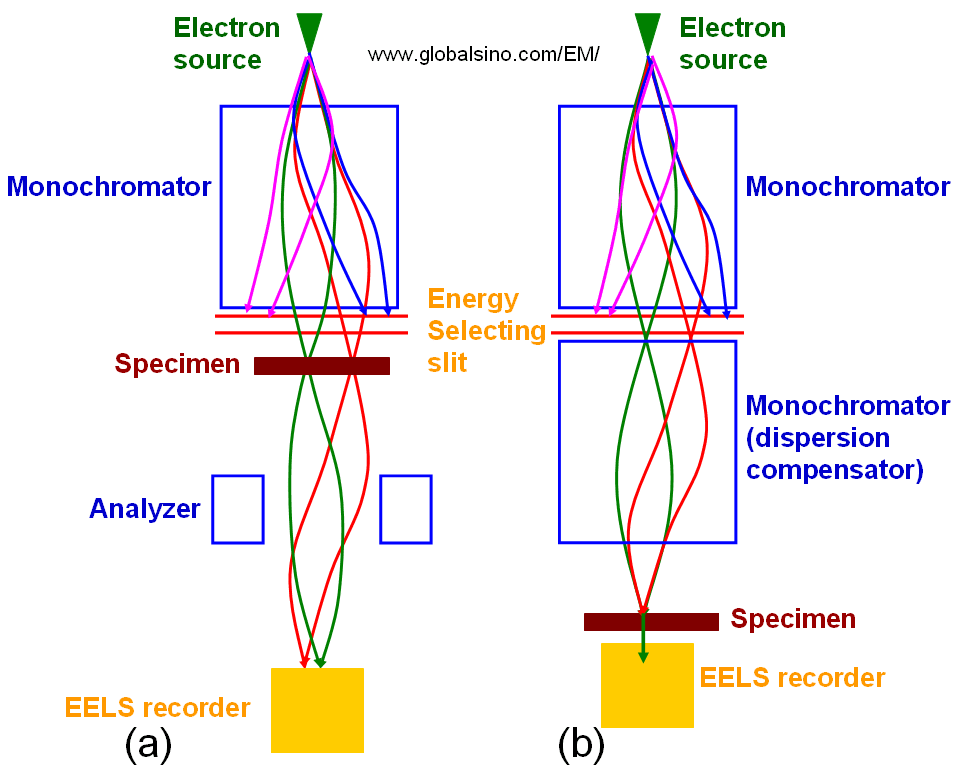
Figure 3641b. (a) Non-compensated EELS, and (b) Dispersion compensated EELS.
[1] Tsuno, K., 2000. Electron optical considerations of a 1 Å STEM with a
monochromator. Microsc. Microanal. 6 (Suppl. 2: Proceedings),
142–143.
|