
Chapter/Index: Introduction | A | B | C | D | E | F | G | H | I | J | K | L | M | N | O | P | Q | R | S | T | U | V | W | X | Y | Z | Appendix
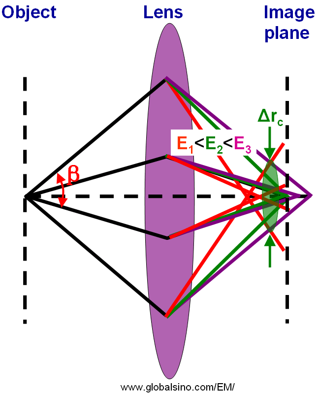
| The instabilities of the accelerating voltage and/or lens currents in the EMs could be contributory factors to the chromatic aberration (Cs), but the two are now negligible because of the high electrical stability of modern power supplies. Even though there is an energy spread (ΔE, 0.3 ~ 1.5 eV) in the electron sources, the major factor to the Cs is the large energy loss ΔE (normally < 2 keV) induced by inelastic scattering when electrons pass through the specimen. Similar to the discussion regarding different accelerating voltages (the focal length at low accelerating voltages is much smaller than that at high accelerating voltages), for chromatic aberration, the light or electrons of different wavelength (or different energy E) are focused differently as shown in Figure 4336. Different color represents different wavelength or energy of rays (light or electrons). There exists a point called the circle of least confusion, where chromatic aberration can be minimized and where the image plane locates.
Figure 4336. Schematic diagram of chromatic aberration. In the case of chromatic aberration, the radius (Δrc) of the circle of least confusion can be given by, Information limit depends on the damping envelope incorporating partial temporal coherence due to chromatic aberration, but not partial spatial coherence due to beam convergence. If we take e-2 as the cut-off value, the information limit due to chromatic aberration is given by [1], where, Δ --- The defocus spread, expressed in terms of the standard deviation.Note that the temporal coherence affects the envelope function because of the energy spread of the electrons, which induces a defocus spread Δ because of the chromatic aberration. The fundamental effect of chromatic aberration (Cc) on TEM imaging is that electrons whose velocities differ from those with nominal velocity are diffracted differently, that is, the slower (longer wavelength) electrons are diffracted more strongly than the faster (shorter wavelength) ones. Round lenses in conventional EMs suffer from spherical aberration as well as off-axial coma. To eliminate the azimuthal or anisotropic coma, the axial magnetic field must change its sign with a dual lens consisting of two spatially separated windings with opposite directions of their currents [2]. The axial chromatic coefficient (Cc) of the coma-free lens is significantly larger (≥ 50%) than that of standard objective lenses. Therefore, in order to obtain sub-Ångstroem resolution it is necessary to greatly minimize the chromatic aberration in coma-free lenses. In HRTEM measurements, the voltage-centering alignment is applied to reduce the image intensity attenuation due to chromatic aberration. In STEM, the fluctuations in accelerating voltage or the current flow in the probe-forming electromagnetic lenses contributes to the chromatic aberration that leads a cutoff to the highest spatial frequency. In HRTEM, the cut-off frequency corresponds to the information limit. Extending Equation 4336a, in STEM, the effect of the fluctuations induces additional contribution to the probe size [3], where, The high-tension power supply and beam-current in modern electron microscopes can achieve 10-7 stability so that the main source of the chromatic aberration is due to energy spread of the electrons within the probe. At high accelerating voltages, the chromatic effect is not as important as the effect of the spherical aberrations as shown in page3644 unless the STEM is operating at a low accelerating voltage such as below a certain voltage depending on the properties of the system, such as 60 kV where the chromatic and spherical aberrations of round lenses are roughly equal. In general, objective pole pieces with small top bore in TEMs have been suggested to have advantages of low spherical and chromatic aberrations and reduced condenser action. Comparing with side-entry specimen stages, top-entry specimen stages have better probe system parameters (e.g. Cs and Cc), and the X-ray spectrometers closer to the specimen.
[1] Spence, J. C. H., Experimental High-Resolution Electron Microscopy, seconded., Oxford University Press, NewYork, 1988.
|
 --------------------- [4336a]
  --------------------- [4336b]
  ------------- [4336c]