Chapter/Index: Introduction | A | B | C | D | E | F | G | H | I | J | K | L | M | N | O | P | Q | R | S | T | U | V | W | X | Y | Z | Appendix
| Table 4310a. Considerations of specimen and microscope parameters in HRTEM imaging.
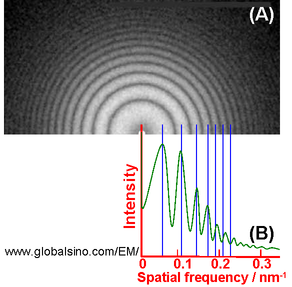
For high resolution TEM imaging, we apply a symmetrical multi-beam illumination. In this case, the transmitted beam and a diffracted beam (e.g. from (hkl) plane) pass through a large objective aperture, and thus interference takes place between the transmitted and diffracted beam, resulting in one-dimensional (1D) lattice fringes of the (hkl) plane. When more diffracted beams together with the transmitted beam pass through, then two-dimensional (2D) high-resolution lattice fringes are obtained. In other words, high-resolution transmission microscopy (HRTEM) uses both the direct electron wave and several diffracted electron waves to form the image. In this way, the resolution is improved with the multiple beams comparing to bright field imaging because higher spatial frequencies are included. Due to their phase differences the interfere between the beams results in an interference pattern in the image plane of the objective lens. Therefore, HRTEM is also a type of phase-contrast microscopy. The coherent low angle elastic scattering can provide information of the relative position of atoms in the TEM specimens and is widely applied in diffraction techniques and HRTEM imaging. In conventional HRTEM, the phase information and amplitude contrast of the exit electron wave are nonlinearly mixed and convoluted with the phase contrast transfer function. This is a rather sophisticated and complex process to obtain the phase and amplitude images. HRTEM usually operates under phase-contrast conditions because the information about the atomic structure of the object consists in the phases of the elastically scattered electron waves. In order to convert a small phase shift into a proper change of contrast, an extra phase shift must be added so that the phase difference between the scattered and unscattered wave is equal to 180°. In conventional TEMs this phase shift is introduced by defocusing the objective lens properly [1]. Thon rings are a phenomenon revealed in the power spectra of micrographs by bright-field (BF) TEM (transmission electron microscopy) imaging. These rings can be explained as the effect of the contrast transfer function, which modulates the Fourier transform of the object in a defocus-dependent way. Figure 4310 (A) shows the power spectrum of a typical BF TEM image of amorphous carbon film presenting concentric Thon rings. Those white rings correspond to the contrast transfer maxima and the dark rings indicate spatial frequency bands without signal. Figure 4310 (B) shows the radial intensity of the power spectra. The astigmatism and defocus can affect the symmetry of the rings, limiting the spatial resolution of the microscope. Therefore, electron micrographs, especially HRTEM, are routinely inspected by optical diffraction before taking images for analysis.
Note again, only electrons scattered at small angles and without loosing energies will contribute to the information in the HRTEM images while all the others (which have lost energy and have been scattered multiple times or at high angles) also contains important information about the sample, they will for instance give a diffuse background in the images and benefit to EFTEM elemental mapping. Furthermore, some aberration coefficients such as spherical aberration, defocus, axial coma, twofold astigmatism, and threefold astigmatism, should be determined for HRTEM imaging at spatial resolutions approaching 0.1 nm or higher. Note that the off-axis aberrations are negligible in the case of high-resolution TEM imaging and STEM [2]. Theoretically, phase extension procedure, using the magnitudes of the structure factors from ED (electron diffraction) and starting phases from the corresponding EM (electron microscopy), can be employed to improve a high-resolution image. Defocus CBED using high-index reflections provides a more accurate determination method of displacement vectors (R) of stacking faults than two-beam method with exciting low-index reflections, selected area electron diffraction method with low index reflections, and HRTEM technique. The diffraction effect of the objective aperture is important for HRTEM imaging. The radius of the Airy disk produced by the objective aperture is given by, Note that the coherence of the electron source is important for high resolution TEM imaging but is not important for all other applications such as Z-contrast STEM imaging, and EDS and EELS measurements. For the same reason, field-emission guns works on HRTEM imaging better than other electron sources (see page4196). HRTEM images can be recorded, for instance, with a Gatan UltraScan 4k×4k CCD camera, and submitted to data processing and analyzed using Gatan DigitalMicrograph software. Table 4310b. Applications of HRTEM technique.
[1] O. Scherzer, J. Appl. Phys. 20 (1949) 20.
|