|
|
History of EELS Technique
- Practical Electron Microscopy and Database -
- An Online Book -
|
|
https://www.globalsino.com/EM/
|
|
|
=================================================================================
Table 3935. History of EELS technique
| Year |
Researcher |
Details |
References |
| Late 1800s |
Starke |
Suggested an electron source could generally distinguish materials of different compositions |
[10] |
| 1904 |
G. Leithäuser |
First attempts of measuring the energy loss of fast electrons traveling through matter date back to the work long before TEM invention
|
[8] |
| 1929 |
E. Rudberg |
Used electron reflection spectrometer to measure energy loss of reflected electrons from copper surfaces. |
[1] |
| 1940s |
James Hillier and R. F. Baker |
EELS technique based on a transmission electron beam was developed. |
[3] |
| 1941 |
G. Ruthemann |
The first measurement of the energy spectra of transmitted electrons using higher incident energy (2–10 keV) |
[5] |
| 1944 |
James Hillier and R. F. Baker |
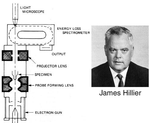
The first study of inner-shell energy loss of transmitted electrons (refer to Figure 3935 below ); first suggested as a micro-analytical technique for elemental detection |
[6] |
| 1948 |
Ruthemann, G. |
Core loss spectrum |
[9] |
| 1960s |
|
Became used practically as a consequence of the progress achieved in coupling well-adapted analyzers and filters to EM columns
|
|
| 1962 |
H. Boersch |
The first Wien filter was used for EELS of transmitted electrons in Berlin |
[7] |
| 1970s |
|
Design of spectrometers and imaging filters flourished |
|
| 1980s |
|
The main use of EELS has been the elemental quantification until the end of the 1980s. |
|
| 1982 |
J. M. Cowley |
Used electron reflection spectrometer to measure energy loss of reflected electrons from materials surfaces in FE-SEM (field-emission SEM). |
[2] |
| 1990s |
|
EELS technique has been widely used in advanced TEM systems at high energy resolutions with high vacuum systems. |
[4] |
| Late 1990s |
|
Schottky emission sources became available and have been providing energy resolution greater than 0.5 eV. |
|
| 2009 |
|
The GIF (Gatan imaging filter) Quantum employs fifth-order aberration correction, a 9-mm entrance aperture, faster CCD readout, and a 1-μs electrostatic shutter to allow simultaneous recording of the low-energy-loss and core-loss regions of a spectrum. |
|
| |
|
|
|
|

Figure 3935. James Hillier (1915-2007) and Baker at the Radio Corporation of America (RCA)
Labs at Princeton, New Jersey, built an electron microprobe, which combined an electron
microscope with an energy-loss spectrometer in 1944.
[1] Rudberg, E. (1930) Characteristic energy losses of electrons scattered from incandescent solids. Proc. R. Soc. Lond. A127, 111–140.
[2] Cowley, J. M. (1982) Surface energies and surface structure of small crystals studies by use of a STEM instrument. Surf. Sci. 114, 587–606.
[3] J. Hillier and Baker, R. F. (September 1944). "Microanalysis by means of electrons". J. Appl. Phys. 15 (9): 663–675.
[4] H. H. Rose "Optics of high-performance electron microscopes - a review" Sci. Technol. Adv. Mater. 9 (2008) 014107.
[5] Ruthemann, G. (1941) Diskrete Energieverluste schneller Elektronen in Festkörpern. Naturwissenschaften 29, 648.
[6] Hillier, J., and Baker, R. F. (1944) Microanalysis by means of electrons. J. Appl. Phys. 15, 663–675.
[7] Boersch, H., Geiger, J., and Hellwig, H. (1962) Steigerung der Auflösung bei der Elektronen-Energieanalyse. Phys. Lett. 3, 64–66.
[8] Leithäuser, G., 1904. Über den geschwindigkeitsverlust, welchen die kathodenstrahlen beim durchgang durch du¨nne metallschichten erleiden, und über die ausmessung magnetischer spektren. Ann. Phys. 15, 283–306.
[9] Ruthemann, G., 1941. Diskrete energieverluste schneller elektronen in festkörpern. Die Naturwissenschaften 29, 648.
[10] Scott VD, Love G (1983) Quantitative electron-probe microanalysis. Wiley, New York.
=================================================================================
|
|
