=================================================================================
The intensity distribution of Fresnel fringes in TEM images varies with the defocus. There is a significant difference between slightly overfocused and underfocused images. Especially, the underfocused image shows an extremely high contrast up to Imax/Imin ~ 3. Here, is the maximum and the minimum intensities.
Generally speaking, spatial resolution in EMs (electron microscopes) is very sensitive to objective astigmatism. Therefore, it is necessary to correct it carefully. In TEM imaging, objective astigmatism is conveniently adjusted by viewing the Fresnel fringes near the edge of a small hole under overfocus condition. If the objective lens field is astigmatic (namely, not cylindrically symmetric), the focus (defocus) will be different along different directions. For instance, those asymmetric Fresnel fringes present around the edge of a round hole in carbon (C) film.
Fresnel diffraction also occurs at the interface between two materials with different densities. At underfocus (the objective lens is focused below the specimen) a bright halo presents outside the area of greater density, while at overfocus (the objective lens is focused above the specimen) a dark fringe appears there. The farther above or below focus the objective lens is, the thicker the Fresnel fringes. At exact focus, no Fresnel fringes are shown and the least amount of contrast in the image is observed.
Figure 4215 shows the TEM images of 1-nm He (helium) bubbles in palladium tritides when the specimen is oriented far from any Bragg conditions of the matrix of the palladium tritides [1]. For negative defocus (underfocus), the bubbles appear as white dots surrounded by a dark fringe, while for positive defocus (overfocus), the dots are black with white fringe.
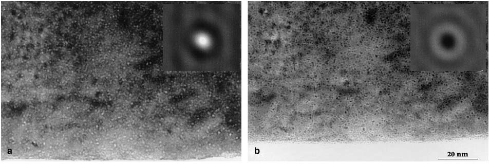
Figure 4215. The TEM images of 1-nm He (helium) bubbles in palladium tritides: (a) Underfocus and (b) Overfocus. The insets present theoretically simulated TEM contrasts of centered He bubbles in 6.5 nm thick TEM specimen. Adapted from [1]
[1] S. Thiébaut, B. Décamps, J. M. Pénisson, B. Limacher, A. Percheron Guégan, TEM study of the aging of palladium-based alloys during tritium storage, Journal of Nuclear Materials 277 (2000) 217-225.
|