=================================================================================
Figure 3526a shows a HRTEM image taken from a cubic perovskite PbMg1/3Nb2/3O3 structure in [111] zone axis [1]. A small deviation from hexagonal symmetry may originate from a slightly tilt away from the exact axial orientation. The inset shows the diffraction pattern of the HRTEM-imaging region.
![HRTEM image taken from a cubic perovskite PbMg1/3Nb2/3O3 structure in [111] zone axis.](image1/3526.jpg)
Figure 3526a. HRTEM image taken from a cubic perovskite PbMg1/3Nb2/3O3 structure in [111] zone axis. [1]
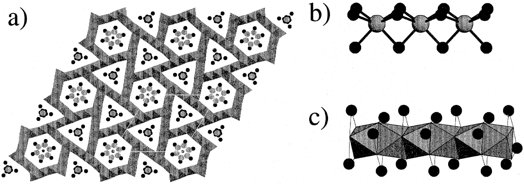
The structure [2] shown in Figure 3526b is formed by a three-dimensional (3-D) Cr7S12 framework raising the strips of edge-sharing octahedra {CrX6} as a triple twinning {111} of NaCl-type and thus, resulting in a hexagonal symmetry sublattice. This 3-D framework forms two different types of tunnels with hexagonal and trigonal geometry. The columns with composition A3CrX3 located in the hexagonal tunnels and the columns with composition A3X located in the trigonal tunnels. There are one hexagonal tunnel and two trigonal tunnels in an unit cell. Both types of columns present hexagonal lattices with the same lattice parameters.

Figure 3526b. (a) Ideal structure of PbCr2S4 projected onto (0001). Pb, Cr and S atoms are drawn as medium black, small black and big gray circles. (b) The triangular tunnels. Black spheres represent Pb atoms and the gray ones located in the center of the face sharing Pb-prisms are S atoms. (c) The hexagonal tunnels. The black spheres represent Pb atoms and the S atoms are located at the corners of the face-sharing octahedra which are centered by Cr atoms. [3]
Figure 3526c shows the SAED (selected-area electron diffraction) patterns of A1-pCr2X4-p crystals along the [0001] zone axis, revealing only the Bragg reflections hk0, which are common to the three sublattices.
![SAED pattern of PbCr2S4 crystals oriented along the [0001] zone axis](image1/3477b.jpg)
Figure 3526c. SAED pattern of PbCr2S4 crystals oriented along the [0001] zone axis. [3]
Surprisingly, the high-resolution images along [0001] (given in Figure 3526d) does not correspond to the expected six-fold symmetry as indicated in Figure 3526c, but to three-fold symmetry.
![HRTEM image of a crystal of a PbCr2S4 crystal imaged along the [0001] zone axis](image1/3477d.jpg)
Figure 3526d. HRTEM image of a crystal of a PbCr2S4 crystal imaged along the [0001] zone axis. The unit cell is outlined. [3]
Table 3526. Some symmetrical diffraction patterns of cubic crystals.
Zone axis |
[100] |
[110] |
[111] |
Symmetry |
Square |
Rectangular |
Hexagonal |
Aspect Ratio |
1:1 |
1: for BCC, SC (almost hexagonal for FCC) for BCC, SC (almost hexagonal for FCC) |
Equilateral |
[1] H. B. Krause, J. M. Cowley, and J. Wheatley, Acta Crystallographica, Section A:
Crystal Physics, Diffraction, Theoretical and General Crystallography, 35, 1015 (1979).
[2] Hyde, B.G., Andersson, S., 1989. Inorganic Crystal Structures. Wiley, New York.
[3] Landa-Cánovas, A.R., Gómez-Herrero, A., and Carlos Otero-Díaz, L. Electron microscopy study of incommensurate modulated structures in misfit ternary chalcogenides, Micron 32 (2001) 481 - 495.
|
![HRTEM image taken from a cubic perovskite PbMg1/3Nb2/3O3 structure in [111] zone axis.](image1/3526.jpg)

![SAED pattern of PbCr2S4 crystals oriented along the [0001] zone axis](image1/3477b.jpg)
![HRTEM image of a crystal of a PbCr2S4 crystal imaged along the [0001] zone axis](image1/3477d.jpg)