Chapter/Index: Introduction | A | B | C | D | E | F | G | H | I | J | K | L | M | N | O | P | Q | R | S | T | U | V | W | X | Y | Z | Appendix
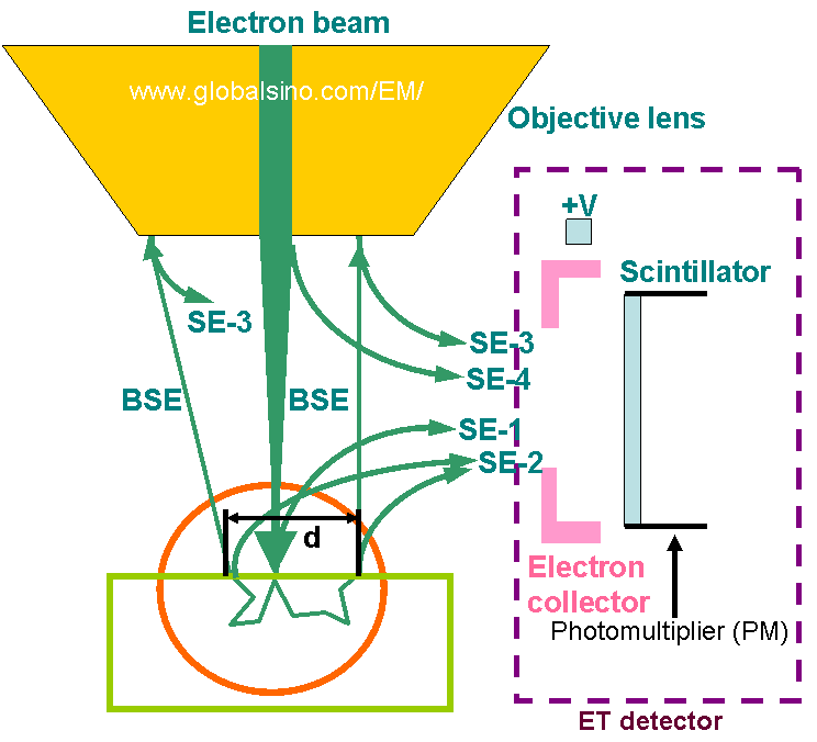
| Most scanning electron microscopes (SEM) images are obtained with Everhart-Thornley (ET or E-T) detectors, as schematically shown in Figure 4584a. This type of detector is named after its inventors, Thomas E. Everhart and Richard F. M. Thornley who published, in 1960, their design to enhance the efficiency of existing secondary electron (SE) detectors. Their main components of the ET detector are a collector grid, a scintillator, and a photomultiplier. They also added a light pipe to carry the photon signal from the scintillator inside the evacuated specimen chamber of the SEM systems to the photomultiplier outside the chamber. The ET detector collects mostly SEs and some BEs (backscattered electrons). The average energy of secondary electrons (SEs) is less than 50 eV and mostly SEs have energies of about 3 to 5 eV. The SEs are pulled toward the scintillator by a high potential (e.g. 300-V) on the collector grid. A high positive bias (e.g. 10-kV) on the scintillator attracts SEs enough to convert SEs to light photons in the light pipe. The collection grid also protects the electron beam from deflection by the 10-kV bias on the scintillator. The ET SE detector can be used for detecting BEs either by turning off Faraday cage or by applying a negative voltage to the Faraday cage. Most of other electrons within the specimen chamber are not pulled and will only reach the detector if their direction of travel takes them to it.
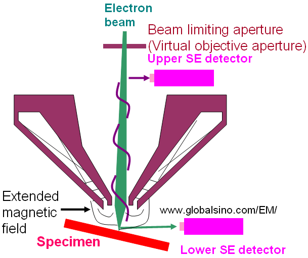
Figure 4584a. Four types of secondary electrons collected by the Everhart-Thornley (ET) detector. Adapted from Reimer [1]. As shown in Figure 4584b, the single-pole snorkel objective lens produces a strong magnetic field that extends from the polepiece directly to the SEM specimen. This type of lenses is a compromise between the aberrations of immersion lens and pinhole lens but accommodate large specimens similar to those in the pinhole lens SEMs. In addition, this configuration can simultaneously accommodate both a conventional (or called lower, e.g. Everhart-Thornley (ET) detector ) and a through-the-lens (or called upper) secondary electron (SE) detectors, providing valuable flexibility for imaging.
Figure 4584b. Single-pole snorkel objective lens.
[1] Reimer, L., Scanning Electron Microscopy, Springer Verlag, New York, 1985.
|