=================================================================================
An advantage with magnetic sector prisms is that they can focus the beam to form a line spectrum in the dispersion plane. In this plane, the electrons are typically dispersed by ~2 µm/eV. Some spectrometers are fitted with a mechanical slit at this plane so that it can be used to select part of the spectrum.
In a EELS and GIF system, a narrow slit is located at the spectrometer image
plane, in front of a single-channel electron detector. Electrons passing through this
slit can be detected in various ways but the most common system uses a scintillator
(to convert the electrons to visible light) followed by a photomultiplier (PM) tube
to produce an electrical signal, e.g Figure 4863a.
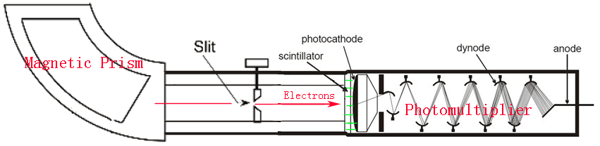
Figure 4863a. Schematic of Serial EELS system.
The electron slit and its surroundings must be carefully designed so as to minimize
the spectrometer background which mainly arises from high-energy electrons
which are backscattered from the slit blades and which, through multiple scattering,
eventually reach the scintillator. For accurate measurements, it is necessary to subtract this background from the acquired data. It is important to make the slit width variable. Gatan EELS systems normally have two slits at different widths. The narrow slit limites spectrometer aberrations and the energy width of
the electron source ( typically 1 – 2 eV) and thus, provides the better
energy resolution, but gives the lower signal counts, therefore, is
more suitable for recording low energy losses. To record inner-shell edges, the wider
slit is needed to obtain data which are not dominated by shot noise.
In parallel EELS
detectors, they contain no energy selecting
slit, so no electrons are wasted, and thus the recording of high energy loss is improved. Figure 4863b shows the schematic illustration of a GIF (Gatan Imaging Filter) camera. The piezo-controlled energy-selecting slit in the GIF system is also indicated in the figure. 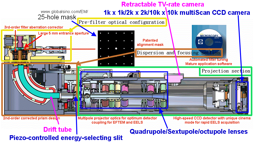
Figure 4863b. Schematic illustration of a GIF (Gatan Imaging Filter) camera. Adapted from [1]
Figure 4863c shows the schematic illustrations of a conventional TEM (a) and a typical omega-filter located between the intermediate lens and projector lens (b). The omega filter is normally about 30 cm in length. The energy selection slit is also indicated in Figure 4863c (b).
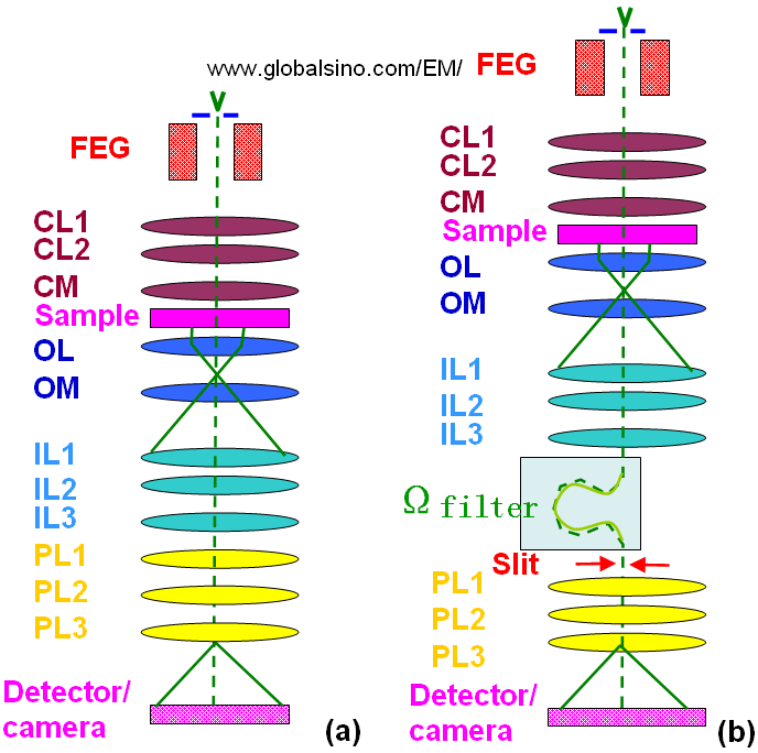
Figure 4863c. Schematic illustration of a conventional TEM (a) and a typical omega-filter (b).
The chemical shift can be mapped by narrow energy slits. For instance, depending on the environment of Si element, the chemical shift of Si L2,3 energy loss edge can be up to 6.9 eV. Therefore, the resulting chemical shift can be mapped using the energy filtered imaging taken with a narrow energy slit of 1.8 eV or similar spectrum mapping in STEM mode.
In energy-filtered TEM (EFTEM) imaging mode, the best to do is that the electron beam energy is increased to preserve image focus. With the “Offset” control on EFTEM interface, the value of the energy loss is chosen. By using the high voltage (tension) offset, an ionization edge can be shifted into focus on slit opening position where the zero loss was located previously. If no slit is applied, the other edges also appear at their corresponding energies but blurred in the vertical direction due to the chromatic aberration.
In operation, the slit may be fully retracted from the beam path by the computer through the EFTEM controller.
There are some methodological differences from system to system, but Figure 4863d shows an example on how the zero-loss-peak alignment is performed. If the beam-pass ratio is at least 25% (or the selected percentage), the zero loss peak of the energy loss spectrum is considered to have been aligned; otherwise, the zero-loss-peak adjustment process is needed. In this system, an upper-slit beam current detector and a lower-slit beam current detector are employed to the detection of misalignment in order to determine whether the majority of the spectrum intensity falls on the upper slit half or on the lower slit half.
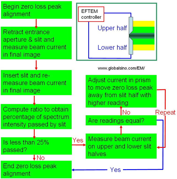
Figure 4863d. An example on how the zero-loss-peak alignment is performed.
The green-boxed inset shows the configuration of the slit.
Spectrum focus adjustment in EFTEM measurement can be done automatically with computer control. Figure 4863e shows an example of adjustment procedures.
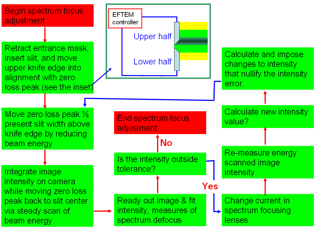
Figure 4863e. Example of procedures of spectrum focus adjustment in EFTEM measurement.
When the energy-loss spectrum is not focused at the energy-selecting slit, normally at low energy-dispersions (high eV/pix values), the shadow of the slit edges in the energy-loss spectrum will not be sharp.
Note that issues from the slit can cause problems:
i) The slit can block the electron beam and then no GIF images can be observed. Therefore, the slit need to be retracted during TEM alignment.
ii) Charging can be induced if the slit is contaminated. In this case, dark and white bands will presents in GIF images (especially in low loss images with small energy-selecting slits).
iii) Slit can block the electron beam if the GIF camera is used to align the electron beam (with Ronchigram) for EELS acquisition in STEM mode.
To know whether the energy-selecting slit is charging or not, we can move the slit edge to the screen under electron-exposure. If the slit is charging, then we may observe:
i) The EEL spectrum or image first gradually drifts in one direction when the charge is building up.
ii) It will suddenly jumps in the opposite direction when the charge dissipates with a discharge.
In a GIF system, if the energy-selecting slit does not move in or out, then we need to check:
i) The presence of pressurized air.
ii) The pneumatic connections.
iii) There should be puffs of air coming from the Gatan Instrumentation Bin (GIB) when the energy-selecting slit is moved in and out in the FilterControl software. In this case, the electronics controlling the pneumatics is working.
iv) Apply a high pressure (≤ 100 N/cm2) to the white slit control hose going into the GIF to loose the o-rings if the GIF has not been used for a long time because the o-rings may stick.
The level of GIF camera chamber vacuum can be evaluated by comparing the current camera chamber vacuum reading to the good ones recorded historically. If the vacuum is poor, one step in the examination procedure to locate any leaks is to observe the vacuum of the camera chamber when you move the energy-selecting slit in/out. It is normal if there is a slight increase in the vacuum reading. However, it also should recover quickly one the movement has stopped. Otherwise, there may be a leak in the seals of the energy-selecting slit.
If the energy-selecting slit is bad or contaminated, then:
i) Spectrum profile and mapping are not affected since the slit is withdrawn in spectroscopy mode.
ii) Lines and (or) bands can exist on EFTEM images when the slit is inserted.
iii) Lines and (or) bands are weak at low loss energies, especially at zero-loss, since the image intensity is strong, but stronger at high loss energies.
iv) The lines and (or) bands are much clear on EFTEM images when a smaller energy window is used.
v) The lines and bands cannot be removed by performing “Remove dark references”.
[1] https://www.gatan.com/.
|