=================================================================================
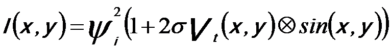
The electron wave undergoes a phase shift, depending on the atomic potential in the specimen, when it penetrates through a TEM specimen. The resulting intensity observed in the image plane of the objective lens is the incident beam intensity modified by this projected potential and an additional term introduced by the imperfections in the lens,
 ------------------ [2765] ------------------ [2765]
where,
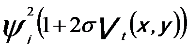 -- The incident wave function multiplied by the projected potential, -- The incident wave function multiplied by the projected potential,
2sin(x,y) -- The lens function, i.e. the effect of lens aberrations on the beam.
HRTEM usually operates under phase-contrast conditions because the information about the atomic structure of the object consists in the phases of the elastically scattered electron waves. In order to convert a small phase shift into a proper change of contrast, an extra phase shift must be added so that the phase difference between the scattered and unscattered wave is equal to 180°. In conventional TEMs this phase shift is introduced by defocusing the objective lens properly [1].
Figure 2765 illustrates the comparison of the main contrasts in both CTEM and STEM modes.
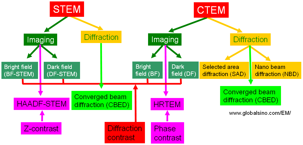
Figure 2765. Comparison of the main contrasts in both CTEM and STEM modes.
However, the phase-contrast term -isin2πχ(g) in the contrast transfer function becomes zero if CS=0 and Δf=0, while the amplitude-contrast term cos2πχ(g) is maximum, 1. Therefore, for aberration-corrected microscope, the TEM images present atomic structures by amplitude-contrast rather than by phase-contrast, and thus in HRTEM images the projected atom column are imaged in bright contrast on a dark background.
[1] O. Scherzer, J. Appl. Phys. 20 (1949) 20.
|