=================================================================================
For high resolution tilt-tomographic imaging in TEMs, the images are recorded every one or two degrees about a tilt axis, over the maximum specimen tilt range. Figure 3983 (a) shows the schematic illustration of tomography imaging with a single tilt holder. The dotted lines represent the imaging sequence taken during specimen-tilting (totally 5 images shown here), while the gray areas indicate the missing wedges of information due to the restricted tilt range. Here, the sample can be too thick or shadowed by the sample holder or other parts of the specimen if conventional TEM specimens and/or specimen holders are used. This tilt maximum results in the wedges of missing information that can induce artifacts. Fortunately, recent development has provided holders to tilt specimens upto 75 -80° that is sufficient to reduce artifacts to a minimum and not lead to serious errors on the measurements of object sizes or shapes.
Figure 3983 (b) shows a so-called dual-axis tomography which reduces the missing wedge. In this way, the tomographic images are recorded with a second tilt series about an axis perpendicular to the axis of the first tilt series. For recording the second tilt series, the specimen is usually rotated by 90°. This dual-axis method with a smaller maximum tilt can improve the quality of the tomographic results as the higher-angle tilts in the case shown in Figure 3983 (a) induce large projected thicknesses.
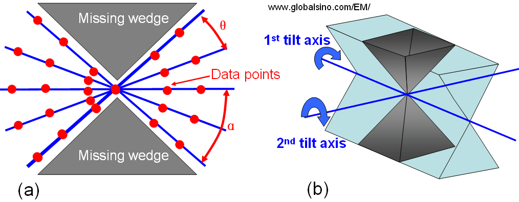
Figure 3983. (a) Schematic illustration showing the tilt sequence during tomographic imaging. θ is the tilt increment between the five images, while α gives the maximum tilt angle.
(b)
Dual-axis electron tomography. The dual-axis tilt series minimize the missing wedge into a missing pyramid of information.
In principle, the resolution of a reconstructed tomogram is determined by the number of images in the tilt series and by the tilt range. In other words, the resolution is equal to the angular increment of the tilt series multiplied by the size of the object [1]. However, in practice, to limit beam damage and to minimize acquisition times, the images are recorded every 1–2°.
Due to the strong interaction of the electron beam with the specimen, bright-field TEM imaging in tilt electron tomography is mainly used in biological tomography but is in general inapplicable to the study of crystalline materials because the diffraction contrast and Fresnel fringes do not satisfy the projection requirement and can lead to serious artifacts in reconstructions. On the other hand, the HAADF (high-angle annular dark-field) STEM imaging offers an excellent alternative due to its Z-contrast.
[1] Crowther, R. A., de Rosier, D. J. and Klug, A. The reconstruction of a
three-dimensional structure from projections and its application to electron
microscopy. Proc. R. Soc. Lond. A 319, 317–340 (1970).
|