Chapter/Index: Introduction | A |
B |
C |
D |
E |
F |
G |
H |
I |
J |
K |
L |
M |
N |
O |
P |
Q |
R |
S |
T |
U |
V |
W |
X |
Y |
Z |
Appendix
Penetration Depth/Implant Depth/Trajectory of Ions in FIB Milling
| Figure 1167 shows the simulations of transport of Ga+ ions near Si (silicon) and Cu (copper) surfaces, indicating the ion trajectories at 0° incidence angle and 88° incidence angle at a 30 keV Ga beam.
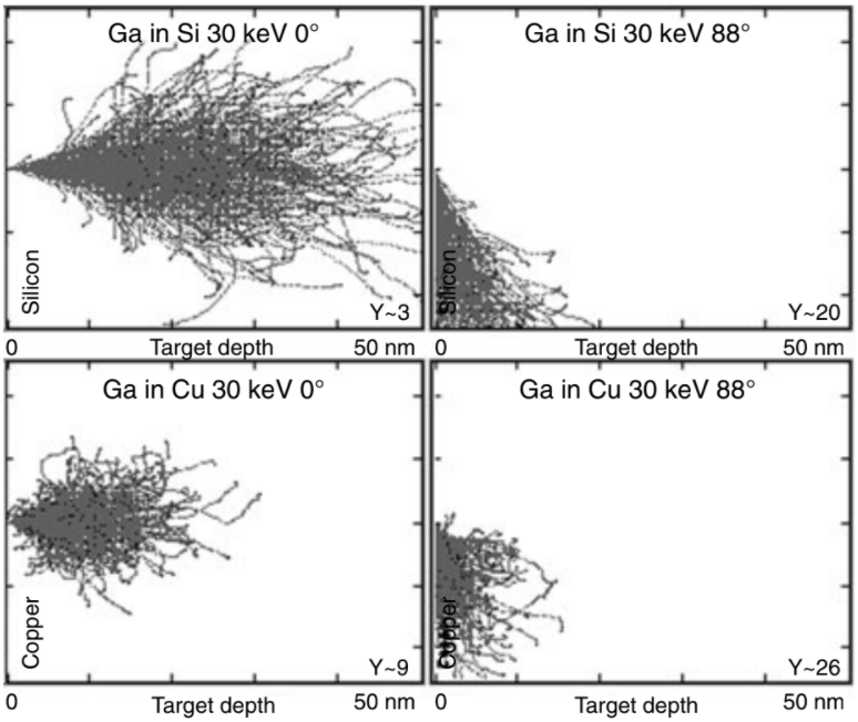
| Figure 1167. Simulations of transport of Ga+ ions near Si (silicon) and Cu (copper) surfaces, indicating the ion trajectories at 0° incidence angle and 88° incidence angle at a 30 keV Ga beam. The full scale for all simulations is 50 nm. The sputter yields, Y, are also given in the figures. [1] |
Table 1167. Penetration depth (implant depth) of ions in FIB milling. |
| Primary ion |
2 kV Ga+ |
|
30 kV Ga+ |
|
|
|
| Angle |
0° |
|
0° |
|
|
|
| Polymer |
12 nm |
|
60 nm |
|
|
|
| 1 |
H |
|
|
|
|
|
|
| 2 |
He |
|
|
|
|
|
|
| 3 |
Li |
|
|
|
|
|
|
| 4 |
Be |
|
|
|
|
|
|
| 5 |
B |
|
|
|
|
|
|
| 6 |
C |
|
|
|
|
|
|
| 7 |
N |
|
|
|
|
|
|
| 8 |
O |
|
|
|
|
|
|
| 9 |
F |
|
|
|
|
|
|
| 10 |
Ne |
|
|
|
|
|
|
| 11 |
Na |
|
|
|
|
|
|
| 12 |
Mg |
|
|
|
|
|
|
| 13 |
Al |
|
|
|
|
|
|
| 14 |
Si |
|
|
50 nm |
|
|
|
| 15 |
P |
|
|
|
|
|
|
| 16 |
S |
|
|
|
|
|
|
| 17 |
Cl |
|
|
|
|
|
|
| 18 |
Ar |
|
|
|
|
|
|
| 19 |
K |
|
|
|
|
|
|
| 20 |
Ca |
|
|
|
|
|
|
| 21 |
Sc |
|
|
|
|
|
|
| 22 |
Ti |
|
|
|
|
|
|
| 23 |
V |
|
|
|
|
|
|
| 24 |
Cr |
|
|
|
|
|
|
| 25 |
Mn |
|
|
|
|
|
|
| 26 |
Fe |
4 nm |
|
20 nm |
|
|
|
| 27 |
Co |
|
|
|
|
|
|
| 28 |
Ni |
|
|
|
|
|
|
| 29 |
Cu |
|
|
25 nm |
|
|
|
| 30 |
Zn |
|
|
|
|
|
|
| 31 |
Ga |
|
|
|
|
|
|
| 32 |
Ge |
|
|
|
|
|
|
| 33 |
As |
|
|
|
|
|
|
| 34 |
Se |
|
|
|
|
|
|
| 35 |
Br |
|
|
|
|
|
|
| 36 |
Kr |
|
|
|
|
|
|
| 37 |
Ru |
|
|
|
|
|
|
| 38 |
Sr |
|
|
|
|
|
|
| 39 |
Y |
|
|
|
|
|
|
| 40 |
Zr |
|
|
|
|
|
|
| 41 |
Nb |
|
|
|
|
|
|
| 42 |
Mo |
|
|
|
|
|
|
| 43 |
Tc |
|
|
|
|
|
|
| 44 |
Ru
|
|
|
|
|
|
|
| 45 |
Rh |
|
|
|
|
|
|
| 46 |
Pd |
|
|
|
|
|
|
| 47 |
Ag |
|
|
|
|
|
|
| 48 |
Cd |
|
|
|
|
|
|
| 49 |
In |
|
|
|
|
|
|
| 50 |
Sn |
|
|
|
|
|
|
| 51 |
Sb |
|
|
|
|
|
|
| 52 |
Te |
|
|
|
|
|
|
| 53 |
I |
|
|
|
|
|
|
| 54 |
Xe |
|
|
|
|
|
|
| 55 |
Cs |
|
|
|
|
|
|
| 56 |
Ba |
|
|
|
|
|
|
| 57 |
La |
|
|
|
|
|
|
| 58 |
Ce |
|
|
|
|
|
|
| 59 |
Pr |
|
|
|
|
|
|
| 60 |
Nd |
|
|
|
|
|
|
| 61 |
Pm |
|
|
|
|
|
|
| 62 |
Sm |
|
|
|
|
|
|
| 63 |
Eu |
|
|
|
|
|
|
| 64 |
Gd |
|
|
|
|
|
|
| 65 |
Tb |
|
|
|
|
|
|
| 66 |
Dy |
|
|
|
|
|
|
| 67 |
Ho |
|
|
|
|
|
|
68
|
Er |
|
|
|
|
|
|
69
|
Tm |
|
|
|
|
|
|
70
|
Yb |
|
|
|
|
|
|
71
|
Lu |
|
|
|
|
|
|
72
|
Hf |
|
|
|
|
|
|
73
|
Ta |
|
|
|
|
|
|
74
|
W |
|
|
|
|
|
|
75
|
Re |
|
|
|
|
|
|
76
|
Os |
|
|
|
|
|
|
77
|
Ir |
|
|
|
|
|
|
78
|
Pt |
|
|
|
|
|
|
79
|
Au |
|
|
|
|
|
|
80
|
Hg |
|
|
|
|
|
|
81
|
Tl |
|
|
|
|
|
|
82
|
Pb |
|
|
|
|
|
|
83
|
Bi |
|
|
|
|
|
|
84
|
Po |
|
|
|
|
|
|
85
|
At |
|
|
|
|
|
|
86
|
Rn |
|
|
|
|
|
|
87
|
Fr |
|
|
|
|
|
|
88
|
Ra |
|
|
|
|
|
|
89
|
Ac |
|
|
|
|
|
|
90
|
Th |
|
|
|
|
|
|
| 91 |
Pa |
|
|
|
|
|
|
| 92 |
U |
|
|
|
|
|
|
| Reference |
|
|
|
|
|
|
[1] Lucille A. Giannuzzi, Particle-Induced X-Ray Analysis Using Focused Ion Beams, SCANNING VOL. 27, 165–169 (2005).
|
