=================================================================================
The image of every electron lens in EMs (electron microscopes) is mathematically formed by a Fourier transformation from the lens front to the back focal plane. That means the spatial coordinates are transformed into their reciprocal values, being spatial frequency spectra theoretically. Those lenses are also called Fourier lens, Fourier transform lens, or Fourier transformation lens, reflecting the two dimensional Fourier transformation takes place. Note that small distances in the object (e.g. between atomic planes) correspond to large spatial frequencies and vice versa.
As shown in Figure 4161a, the electrons propagate from the object as a spherical wave and cross the two lenses in the set-up sequence, appearing in the 1st and 2nd Fourier Transform Planes.
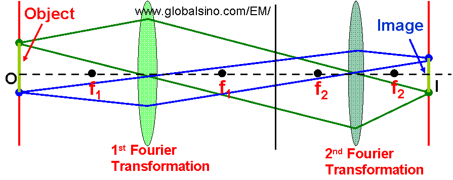
Figure 4161a. Fourier transformation lens in EMs
For an ideal objective lens, the incident electron probe simply forms an Airy disc in the back focal plane of the lens. This disc is the Fourier transform of the uniformly illuminated condenser aperture.
|