Chapter/Index: Introduction | A | B | C | D | E | F | G | H | I | J | K | L | M | N | O | P | Q | R | S | T | U | V | W | X | Y | Z | Appendix
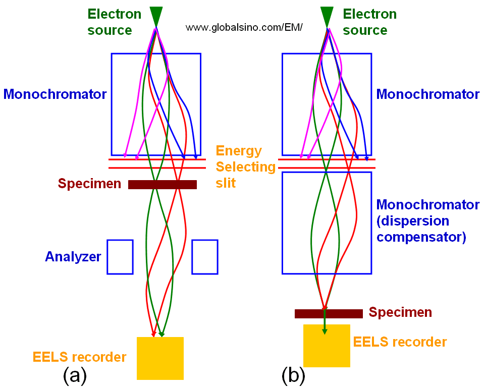
| In order to improve the energy spread of the electron sources in EMs, monochromators have recently been introduced. In the best cases, the energy spread with a monochromator for any source can be reduced to < ~100 meV. There are two basic setups: the Wien filter with crossed electric and magnetic fields and the electrostatic Omega filter. It is very common that both types improve the energy width to about 0.2 eV at an acceptable beam current of several 100 pA. The single Wien filter limits the probe size to about 2 nm, whereas there is no such limitation in the case of the Omega filter. This spatial aberration of the Wien filter can be improved by using double Wien filters [1]. In general, the popular choice is the Wien filter. In ideal EMs (electron microscopies), e.g. in point-like monochromatic TEM (transmission electron microscopy), we can assume that the electron beam is generated from a point-like source and the energy variation ΔE is zero. This electron source is fully coherent. Even though the TEM specimen can be a crystal and thus, the incident electron beam k0 is split into different Bragg-diffracted components kg, resulting in a characteristic phase shift, the phase relation between the diffracted beams is maintained (the elastic electron scattering is coherent) [2]. This coherent relationship induces an interference pattern forming a HRTEM image. In advanced STEM-EELS systems, the monochromators induce an energy spectrum of the electron source at a dispersion plane, where a small fraction of the electron distribution is selected by a narrow energy-selecting slit for the measurements as shown in Figure 4194. A drawback of this method is that the electron beam width is increased in the dispersion direction as shown in Figure 4194 (a). In some high-dispersion designs [3], this large dispersion is compensated and the electron beam is reduced to its original size by another monochromator which produces an achromatic image of the electron source as shown in Figure 4194 (b).
Figure 4194. (a) Non-compensated EELS, and (b) Dispersion compensated EELS.
[1] Martínez G, Tsuno K. 2004. Design of Wien filters with high resolution. Ultramicroscopy 100:105–14.
|