Chapter/Index: Introduction | A |
B |
C |
D |
E |
F |
G |
H |
I |
J |
K |
L |
M |
N |
O |
P |
Q |
R |
S |
T |
U |
V |
W |
X |
Y |
Z |
Appendix
Astigmatism Correction Coil Control System in SEMs
| Astigmatism is a common issue in Scanning Electron Microscopes (SEMs) that arises when the electron beam does not converge into a single focal point, leading to image distortion. This distortion occurs because the electron beam's shape becomes elliptical rather than perfectly circular as it interacts with the lenses. The result is a loss of resolution and clarity in the SEM images, which can reduce accurate analysis. Astigmatism in SEMs is typically caused by imperfections or asymmetries in the electron optics, for instance:
- Lens Imperfections: Slight asymmetries in the electromagnetic lenses that focus the electron beam.
- Contamination or Damage: Particles or damage on the lens surfaces can create uneven electric or magnetic fields, distorting the beam.
- Mechanical Misalignments: Misalignments in the lens system or other components of the SEM can introduce astigmatism.
To correct astigmatism, SEMs are equipped with stigmators, which are specialized electromagnetic coils positioned in the electron optics system. These coils generate an adjustable magnetic field that compensates for the asymmetries causing the beam distortion. The stigmators can correct the beam shape by adjusting the field to restore the beam to a more circular cross-section as it passes through the focal point. The astigmatism correction coil control system in SEMs involves several key components:
- Stigmator Coils: These are the primary hardware elements that generate the corrective magnetic field. Typically, there are two pairs of stigmator coils (one pair for X-axis correction and another for Y-axis correction).
- Control Electronics: The control electronics regulate the current supplied to the stigmator coils, thereby controlling the strength and orientation of the corrective magnetic field. Precise control is essential, as the field must be finely tuned to counteract the specific astigmatism present in the system.
- User Interface: Modern SEMs provide an interface that allows the operator to manually adjust the stigmator settings or use automated correction routines. These routines often involve the SEM software analyzing the beam shape and making iterative adjustments to the stigmator coils until optimal focus is achieved.
- Feedback Mechanisms: In some advanced SEMs, feedback systems are used to continuously monitor the beam shape and automatically adjust the stigmators to maintain focus as conditions change (e.g., during long imaging sessions or when the specimen is moved).
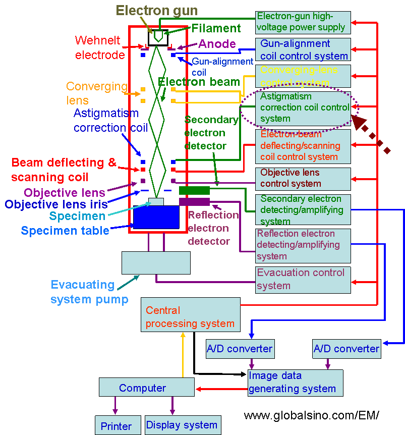
Figure 4226 shows an example of SEM systems. In all EMs (electron microscopes), the converged electron beam passes through the astigmatism correction coil, which is controlled by an astigmatism-correction coil control system, to control a beam shape.

Figure 4226. Example of computer-controlled EMs: SEM system.
|
