Chapter/Index: Introduction | A | B | C | D | E | F | G | H | I | J | K | L | M | N | O | P | Q | R | S | T | U | V | W | X | Y | Z | Appendix
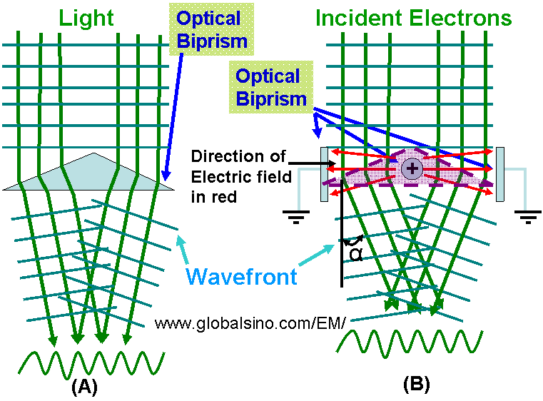
| Figure 4307 (A) shows the schematic illustration of an optical biprism. Similar to the optical biprism, Möllenstedt–Düker biprism [1] consists of a positively charged wire placed between two grounded plates as shown in Figure 4307 (B). The distribution of the electric fields around the charged wire are not uniform because of the difference between the wire and the plate sizes. Note that the violet biprism in (B) does not physically exist but reflects the function of the nonuniform electric fields. The deflection angle (α) of the electrons is proportional to the voltage Ubp between the filament and the plates, α=A0Ubp ------------------ [4307] with the deflection coefficient (A0) dependent on biprism dimensions.
Figure 4307. Schematic illustration of Möllenstedt–Düker biprisms in optical and electron microscopes.
[1] Möllenstedt, G. and Düker, H., 1956. Beobachtungen und messungen an biprisma-interferenzen mit elektronenwellen. Z. Physik 145, 377-397.
|