Chapter/Index: Introduction | A | B | C | D | E | F | G | H | I | J | K | L | M | N | O | P | Q | R | S | T | U | V | W | X | Y | Z | Appendix
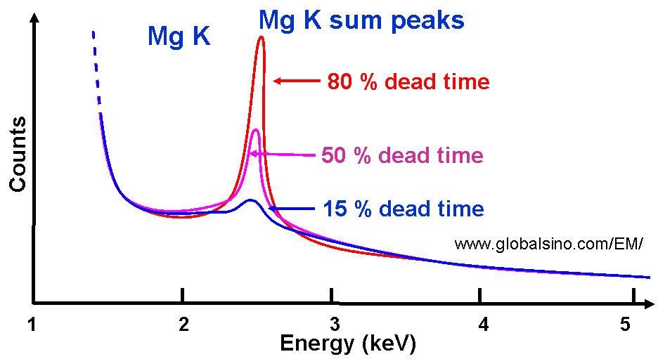
| The EDS data collection is done in a pulse form. When the incoming voltage or current pulses are measured by a pulse processor in the EDS detector, the detector is effectively switched off for the period of time termed as the dead time. In other words, when the EDS system is not counting incoming X-ray photons but processing the previously collected signal, it is said to be ‘dead’. The dead time is given by Dead time in % = (1-Rout/Rin) × 100% ------------------------------- [4639] where, In general it is preferred to have an appropriate value of the dead time because one challenge in operating an EDS system is ensuring that enough X-ray emissions are counted and plotted to come up with a valid EDS spectrum. A high dead time means the detector is swamped with X-rays while the collection becomes inefficient (Rin is too low as indicated in Equation [4639]). In this case, the EDS spectrum can have no peaks. The dead time can be very high if an objective aperture is in the EM (electron microscope) column because it generates X-rays. On the other hand, Brightness and Spot Size functions in EMs can also affect the dead time. Generally speaking, the good dead time for data acquisition is in the range of 15 to 40 % (or alternatively at input count rates between ~1500 and 5000 cps) depending on the specific EDS and electron microscope. Therefore, the input count rate of EDS detector is normally selected so that the system deadtime is generally less than 30%-50% in order to minimize the effects of pulse pileup and peak distortion. However, when minor and/or trace elements are of interest, a high deadtime, e.g. 30–40%, should still be used to record the spectrum regardless of the artifacts mentioned. To further verify the EDS acquisition system, the dead time should be lower than 2% if the electron is moved into a hole (vacuum). Note that a higher dead time value will be obtained if the EM system, including apertures and spot size, is not set correctly. Figure 4639 shows the schematic illustration of dependence of sum peak intensities on different dead times. This figure indicates that we need to minimize the dead time in order to minimize the intensity of the sum peak, especially when the sum peak overlaps the original characteristic x-ray peaks from the EM sample.
Figure 4639. Schematic illustration of dependence of sum peak intensities on different dead times. Table 4639 shows the effect of processing time on spectral parameters. The peak width determines the energy resolution of the spectrum. Due to limitations of current digital pulse processing systems, at high deadtime, EDS spectra are subject to significant pulse coincidence, resulting in pile-up peaks as well as pile-up distortions to the background. Table 4639. Effect of processing time on spectral parameters.
|